

Guided surgery with immediate loading report: a case report
Authors: F. Zingari F. Gallo S. Ghezzi E. Carozzi

A 63-year-old female patient, smoker, in good general health, with severely compromised dental arches.
X-ray examination with orthopantomography showed severe periodontal disease in the few remaining teeth in the arches and significant bone resorption in the maxilla, resulting in a severe loss of vertical dimension. Cone Beam CT is required in order to establish a proper treatment plan. (PICTURE 1,2)

picture 1

picture 2
TREATMENT PLAN
Once CT scan is viewed it was possible to establish the treatment plan that provides, first of all, the rehabilitation of the vertical height through the insertion of a temporary upper and lower prosthesis anchored to the elements still present in the arch, less compromised, elements 12 and 11 will be extracted. Then the bone graft will be performed in the upper arch, with large bilateral maxillary sinus elevation. Subsequently, after removal of the remaining teeth, 7 implants will be placed in the upper jaw and 8 in the lower jaw with immediate loading in guided surgery.
PRE-SURGICAL PHASE
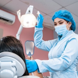
The therapeutic project begins with the taking of impressions with an intraoral scanner and several photos of the patient in front and in profile with mouth half-closed and smiling, which will be necessary for the realization of first two prototypes and then the first temporary prosthesis. (PICTURE 3)

picture 3
These will allow to re-establish a correct vertical dimension and to make all the necessary aesthetic evaluations and adjustments to arrive at immediate loading with a new temporary prosthesis, which will satisfy both, the aesthetic and functional features.
These temporary prosthesis will adopereted in the first phase of treatment so that they can be functionalized for a correct time, given the increase in vertical dimension introduced.
After a CT scan of the maxillary sinuses, necessary to check the absence of sinusitis and the perviousness of the ostium-meatal complexes, the patient is ready to face the first surgical phase.
SURGICAL PHASE
The first surgical phase involves bone grafting in the upper arch by means of a large bilateral maxillary sinus lift, to compensate for the severe atrophy present.
The surgical act involves a paramarginal incision in the vestibular region 14-17 and 24-27, skeletonization and ostectomy in the lateral maxillary wall, elevation of Schneider's membrane and its reinforcement with a PRF membrane, insertion of the bone graft and application of a another membrane to close.
The patient will wait 6 months for complete healing of the surgical site before continuing with the implant surgical phase.
Once the necessary time has passed, the patient undergoes new in-traoral scans, necessary for the creation of a stent, an extraoral geometry that the patient will in-place during the CT scan and that allows for the realignment of the files during the digital project phase.
At this point the files of the intraoral scan and the CBCT are coupled and allow to realize a digital diagnostic wax-up and to decide the best implant position, analyzing quality and quantity of bone in relation to the future position of the teeth. In this way an individualized implant-prosthetic project is obtained.
The second surgical phase involves, therefore, the remaining teeth extraction in the upper and lower arch, the positioning of the implant surgical guide and the insertion of 7 implants and 4 mini implants in the upper arch and 8 implants in the lower arch.
Guided surgery requires all steps to be guided by indexed sleeves in which the drills can be uniquely positioned in a three-dimensional sense with a working length stop, and also requires that the implant be inserted through the same sleeves without removing the template. The sequence of drills that is used is methodically adhered to in order to achieve a predictable result.
In both arches an immediate load is realized; in the upper arch in particular, however, this is possible by using the mini implants, as the quality of the bone did not allow for primary stability and sufficient torque (35N) to load the implants. Therefore, we will wait 4-6 months and then perform a traditional loading. (PICTURE 4)

picture 4
PROSTHETIC PHASE
Thanks to the implant-prosthetic design carried out in the pre-surgical phase, it is possible to place the temporaries in the same session as the surgery, immediately regaining a correct occlusion, vertical dimension and satisfactory aesthetics. Once 4 months have passed after implant surgery and the implant sites and soft tissues have healed completely, we are ready to proceed with the definitive prosthetic phase.
First of all, the implants in the upper arch that could not be possible immediately load, are uncovered, and then new impressions are taken with an intraoral scanner that will give the laboratory the exact position of all the implants and the new state of the soft tissues.
Once we have the new temporary prostehesis, it is possible to remove the mini implants, and proceed with the production of new prototypes with which we will make the final evaluations and modifications. (PICTURE 5)

Before finalizing the restoration, a cobalt-chrome bar is designed by CAD and then tested in the mouth on the turrets to check the passivation, also radiographically. (PICTURE 6)

Once all the esthetic and functional requirements have been met, we are ready to proceed with the production of the final monolithic zirconia restorations, with which the last esthetic tests are performed before the final positioning in the mouth. (PICTURE 7).
The angled screws are tightened to 25N and the straight ones to 35N. On element 13, to hide the vestibular presence of the turret through-hole, a disilicate veneer was created and cemented in the site. (PICTURE 8)

picture 7

picture 8
 Related articles

The aim of the present study was to conduct a critical literature review about the technique of computer-guided surgery in implantology to highlight the indications, purposes, immediate loading of...
News 22 September 2023

The new software aims to streamline the guided surgery workflow and provide dentists with a comprehensive guided surgery solution.
USA 09 June 2023 - 11 June 2023
Freehand vs. guided: from start to finish - 2023 AAID Central District Meeting

The program will cover the many challenges with free-hand and guided surgery in patients with inadequate bone.
Prosthodontics 23 March 2023
Video: Dr. Nick Fahey, author of “Guided Surgery: Making Implant Placement Simpler”

Dentistry is a hard and rewarding career, Fahey added. “With technology and the things available to us, it’s an incredibly interesting career option,” he said.
Implantology 23 March 2023
Dr. Yaste's new book, “Pragmatic AOX,” highlights the use of a stackable surgical guide systems to perform full-arch implantology and rehabilitation.
Read more
Products 26 June 2026
Coronal flaring is considered a key step in efficient root canal preparation, especially for difficult-to-access canals.
News 26 June 2026
The Association for Dental Safety (ADS) proudly announced the recipients of the 2026 Leadership Awards during its Annual Conference on May 27 in Salt Lake City, Utah.
News 26 June 2026

Recognition highlights the company’s doctor-led culture and continued investment in team member growth and engagement
Editorials 26 June 2026
From California to Canada: CU Anschutz School of Dental Medicine at ADEA, GRC and IADR 2026
The CU Anschutz School of Dental Medicine will showcase a wide breadth of educational innovation, faculty development, clinical training and research at three major gatherings this
Oral surgery 26 June 2026
This peer-reviewed oral surgery article summarizes clinical evidence from International journal of oral and maxillofacial surgery (2026). It focuses on findings that may help dental professionals...


Copyright © 2026 - All Rights Reserved
ISSN 2767-1178