

Endodontic instruments fractured in the canals: how to locate them?
Simona Chirico
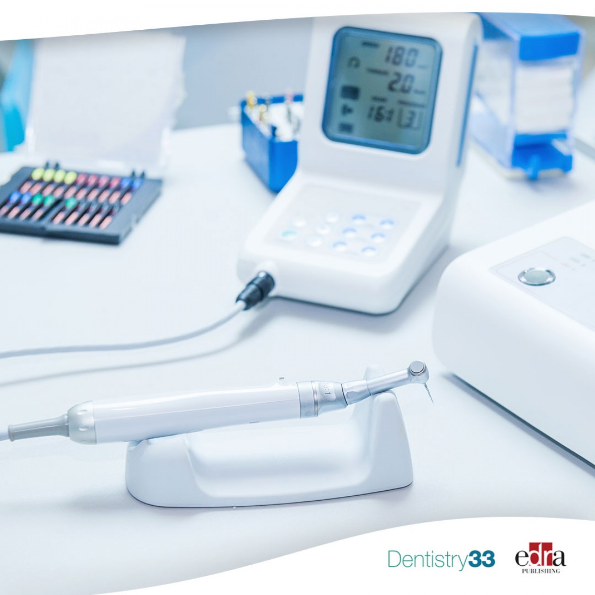
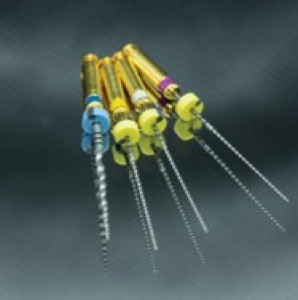
Fracture of shaping instruments within root canals is a common undesirable incident in endodontics. These fractures can prevent proper preparation, disinfection and obturation of the canal. They can also cause significant sequelae on the outcome of endodontic treatment and have an impact on treatment planning. In addition, there are potential legal ramifications.
Radiographic evaluation is a crucial step in the management of endodontic complications but can be essential for the identification of fractured root canal instruments within the canals.
Conventional and digital periapical radiography (PAR) provide limited information due to the conversion of three-dimensional (3D) anatomy into a two-dimensional (2D) image. Therefore, the overlapping of anatomical structures may reduce its diagnostic efficacy.
Cone beam computed tomography (CBCT) allows for 3D evaluation of teeth and surrounding anatomical structures. In endodontics, CBCT is used to evaluate complications such as fractured instruments within root canals but has limitations, including higher patient radiation exposure than PAR.
Materials and methods
In a study published in Oral Surgery, Oral Medicine, Oral Pathology and Oral Radiology in September 2022, the authors compared the diagnostic efficacy of periapical radiography (PAR) with that of CBCT. They analyzed the effect with and without a metal artifact reduction algorithm (MARA) in the detection of fractured endodontic instruments, in plugged and non-obturated, straight and curvilinear canals.
In total, 144 root canals of 48 mandibular molars were divided into four groups:
- Group 1 or control group with empty channels
- Group 2 or fracture group with fractured instrument in canal
- Group 3 or filling group with root canal filling, and
- Group 4 or fracture/fill group with a fractured instrument and root canal filling.
Teeth were radiographed by PAR using a complementary metal oxide semiconductor sensor and by CBCT without and with MARA to evaluate the diagnostic efficacy of the three techniques in detecting fractured instruments. Three examiners evaluated the radiographs.
Results
Overall, PAR showed significantly better diagnostic outcomes than CBCT without MARA. PAR produced significantly better results than both CBCT protocols in the presence of root filling and in straight canals. In the absence of filling in the curved canals, no statistically significant differences were found between the 3 techniques.
Conclusions
Periapical radiography is the imaging technique that best detects fractured instruments in straight obturated and non-obturated root canals.
However, researchers found no significant differences between the three imaging modalities in detecting fractured files in the absence of filling material in curved canals.
CBCT with MARA has shown greater sensitivity and specificity for the detection of fractured instruments in curved canals compared to straight canals.
For more information: "Diagnostic efficacy of three imaging modalities in the detection of fractured endodontic instruments: an in vitro study."
 Related articles
Endodontics 17 February 2026
Treatment options for endodontic failure include nonsurgical or surgical endodontic retreatment, intentional replantation, and extraction with or without replacement of the tooth.
Digital Dentistry 09 June 2023
Association between intracanal medicament radiopacity, streak artifact production using CBCT

This study aimed to calculate the correlation between the radiopacity levels of various intracanal medicaments and radiolucent streak formation using cone-beam computed tomography.

This meta-analysis sought to identify the in vivo prevalence and influencing factors of middle mesial canal in mandibular first and second molars based on cone-beam computed tomography scans.

A study in the Angle Orthodontist evaluated the validity and reliability of marginal bone level measurements made on CBCT images produced using two reconstruction techniques, two viewing modes, and...
Endodontics 28 March 2023
External cervical resorption: relationships between classification, treatment, and outcomes

The purpose of this study was to identify possible associations between classification, treatment, and one-year outcome of external cervical resorption (ECR) lesions using the Heithersay and Patel...
Read more
Eagle Crown Lengthening Burs are designed to make surgical precision effortless—helping clinicians expose more tooth structure smoothly, efficiently, and with total control.
News 17 July 2026
The new Bogotá, Colombia facility strengthens Roland DGA’s long-standing commitment to dental professionals and partners across the region.

New integration streamlines patient financing within CareStack’s practice management platform, making it easier for providers to help patients move forward with care.

As the University of Colorado School of Dental Medicine celebrates the graduating DDS Class of 2025, we are proud to recognize the students and faculty members whose exceptional de
Oral surgery 17 July 2026

This peer-reviewed oral surgery article summarizes clinical evidence from International journal of oral and maxillofacial surgery (2025). It focuses on findings that may help dental professionals...


Copyright © 2026 - All Rights Reserved
ISSN 2767-1178