


Minimally invasive restoration with ultrathin veneers and posterior partial restorations
By Dr. Francisco García Torres and Alexander Lichtmannegger

The article presents a case made for a young patient with generic dental wear, loss of vertical dimension, interference in the posterior area and absence of canine and anterior guidance. Treatment included partial zirconia restorations, a three-unit bridge and ultra-thin zirconia veneers. The veneers are produced with a new technique that involves minimal or no preparation of the tooth (Prettau® Skin®) and the use of individual preparation guides.
On clinical examination, clinicians found the young patient had generic dental wear, with loss of vertical dimension, interference in the posterior area and absence of canine and incisive guidance.

In the light of what was observed, the clinician decided to treat the case by adopting a minimally invasive approach, with a treatment that consisted of: ultra-thin veneers made according to the Prettau Skin® technique in areas 13-23 and 33-43; posterior partial restorations in areas 24, 25, 14, 15, 34, 44, 45 (vonlay) and 16, 17, 26 and 27 (table-top); a minimally invasive partial bridge bonded with vonlay and table-tops in zones 35, 36 and 37.
The treatment was conducted digitally, starting from the diagnostic phase with the use of the PlaneSystem (M. Odt Udo Plaster, Germany) and the 3D Face Hunter facial scanner.
These tools allowed the virtual 3D reproduction of the patient's face and oral situation, considering any occlusal asymmetries between the arches.
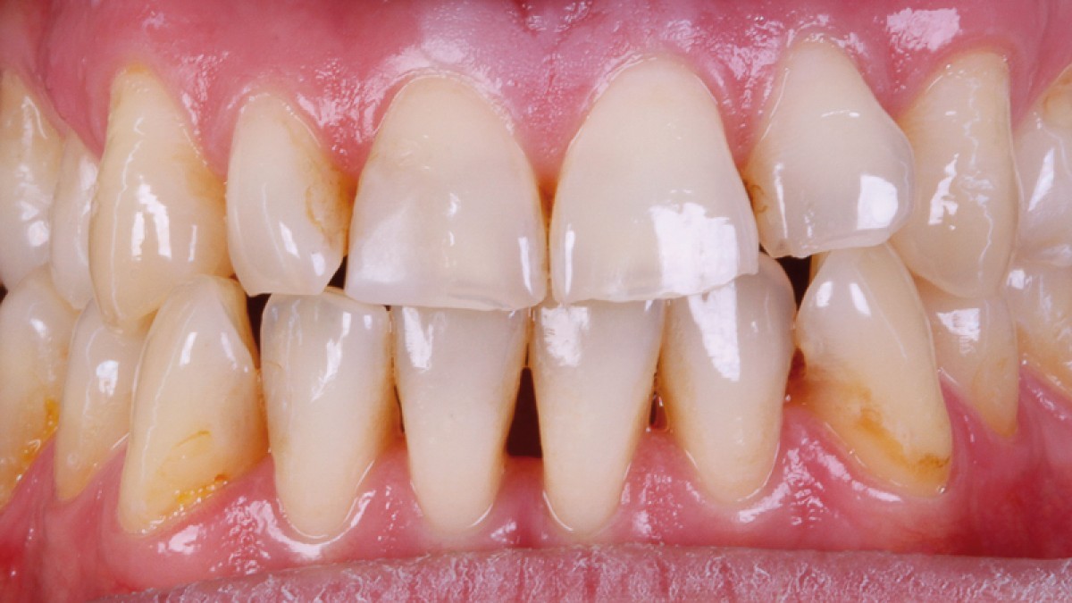
With the aim of restoring correct canine and anterior guidance, the clinician also eliminated the present occlusal interferences by increasing the vertical dimension and reconstructed the areas subject to functional wear.
The Zirkonzahn Shade Guide Prettau Line shade guide was used to choose the most suitable shade of zirconia.
Prettau 2 Dispersive (A1) zirconia was selected for this treatment, a material that combines high flexural strength with excellent translucency and is already equipped, during production, with a natural color gradient that makes the optional ceramic coating.
The teeth were set up with the Zirkonzahn.Modifier software, in which the Heroes Collection dental anatomy library is available. The dental technician selected the most suitable morphology from the library, which he subsequently individualized.
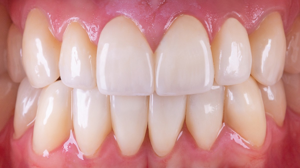
After the try-in of the prototypes, the dental technician designed and created a patient-specific preparation guide for the dentist, marking the areas of the teeth to be prepared for the application of the veneers. This preparation mock-up is a useful tool to optimize planning and communication between dental technician and dentist, with the goal of providing the best possible patient care.
Before starting the treatment and applying the definitive material, dentists conducted prophylaxis and removed all carious lesions. After that, the clinician proceeded with the minimally invasive preparation of the teeth using the guides received, which allowed optimal control of the reduction of the dental tissue.
All necessary adjustments were made using fine diamond burs and finishing and polishing discs.
Subsequently, the clinician took the impression and sent it to the dental technician for modeling of the final restoration. After milling in Prettau 2 Dispersive zirconia and sintering, the restoration was characterized with ICE Stains 3D by Enrico Steger for an even more personal aesthetic result.
The 0.2 mm thick veneers were created using the Prettau Skin technique developed by Zirkonzahn. This method allows patients to be given a new aesthetic smile with no or minimal preparation of the dental substance and is indicated for the aesthetic correction of dental dyschromia, irregular interdental spaces, crooked, malformed or abraded teeth.
The restorations were bonded after treatment of the tooth tissue surface (total etching of the enamel in three different steps) and application of 37% orthophosphoric acid for 20 seconds, chlorhexidine to inhibit metalloproteins, primer and OptiBondTM FL adhesive (Kerr) on the surface of the enamel, all followed by the polymerization phase.
The inner surface of the zirconia was treated following the Zirkonzahn protocol (sandblasting with aluminum oxide, 100 micron at 2 bar, application of Tokuyama universal primer and Estecem II cement).
After the excess material was removed, final polishing was performed.
Dr. García Torres is based in Mexico and Alexander Lichtmannegger is a dental technician and educator with Zirkonzahn Education Center in Bruneck, Italy.
 Related articles
Implantology 03 February 2026
Bone Structure, Metabolism, and Physiology Its Impact on Dental Implantology
When placing implants in the mandible or maxilla, it is important for clinicians to understand the process of bone remodeling, the different types of bone, and how these factors can affect the...
Endodontics 02 February 2026
Implantology 20 January 2026
Smoking as a risk factor for dental implants and implant-related surgery

Cigarette smoking is known to adversely affect wound healing, and thus may jeopardize the success of dental implantation and implant-related oral surgery.
Products 27 November 2025
Neocis Unveils Next-Generation AI-Powered Robotic System for Dental Implants

Neocis, the pioneer behind the first and only U.S. FDA-cleared robotic system for dental implant surgery, today announced the launch and FDA approval of its next-generation robotic platform: Yomi...
Read more
Products 15 October 2026
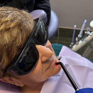
The belief in the healing power of light goes all the way back to ancient Egypt and the worship of Rah, the sun god.
Nearly half the world’s population is affected by oral disease—and the number of cases is rising.
News 15 October 2026

New office at 3170 R Street adds walk-in availability and same-day care options in a region where dental providers are in short supply
Each year, the University of Colorado Anschutz School of Dental Medicine (SDM) Excellence Awards recognize individuals whose dedication, leadership and innovation strengthen our mi
Oral surgery 15 October 2026
This peer-reviewed oral surgery article summarizes clinical evidence from International journal of oral and maxillofacial surgery (2026). It focuses on findings that may help dental professionals...


Copyright © 2026 - All Rights Reserved
ISSN 2767-1178