Limitations and factors influencing molar distalization
Davide Elsido
Molar distalization can relieve dental crowding when neither arch expansion nor labial inclination can obtain a stable space for non-extraction orthodontic treatment. It involves moving molars into the posterior region of the alveolar bone, yet molars can be distalized only as far as there is an effective bony envelope to house the roots.
The success of such treatment depends strongly on the clinician’s thorough understanding of the topography of the region, exceeding which will result in periodontal complications such as dehiscence, orthodontically induced root resorption and tooth mobility. The boundaries of the posterior alveolar region are an important factor to consider in the treatment planning of molar distalization.
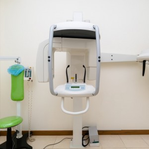
In the October 2022 issue of Angle Orthodontist, a group of researchers from China published a study to analyze the anatomical limitations and characteristics of maxillary and mandibular retromolar regions affecting molar distalization using cone-beam computed tomography (CBCT).
A total of 120 patients were classified into equal groups of skeletal Class II and Class III and stratified by vertical growth pattern, age, sex and third molar presence. The available distance along the axis of distalization and cortical bone thickness (CBT) were measured in the maxillary and mandibular retromolar regions of Class II and Class III patients, respectively. One-way analysis of variance was used to examine the effects of the factors on the measured data.
A posterior occlusal line (POL) connecting the buccal cusps of the first and second molars at the occlusal level was drawn. This represented the axis of distalization (AOD) along the dental arch.
The distance between the root of the second molar and inner cortex of the bony envelope directly reflects the available area where the tooth is allowed to move along the AOD within the bony envelope before the root encounters cortical resistance. Attempts to distalize molars into a retromolar region that cannot fully envelope its roots will result in many complications.
The results suggested that the anatomical limits of molar distalization can be evaluated on the axial slices of CBCT images, along the AOD. The available distance in the retromolar region of the Class II maxilla increased apically and hence should be observed at a limit level closest to the occlusal level. Conversely, the available distance in the retromolar region of the Class III mandible decreased apically and should be observed at a level closest to the apical level. The CBT observed at the limit levels represented the loci where the distally moving molars reach the inner cortex of cortical bone, exceeding which will result in periodontal complications.
Previous studies have demonstrated that hyperdivergent patients had thinner alveolar ridges and were more susceptible to periodontal-related iatrogenic complications, because there is less effective space for the teeth to move.
Researchers observed that the available distance along the AOD showed decreasing trends from hypodivergent to normodivergent to hyperdivergent groups successively in both the Class II maxilla and Class III mandible. This trend was especially significant in the Class III mandible, signaling that mandibular molar distalization is especially risky in the hyperdivergent Class III patient.
The influence of vertical growth patterns on the structure of the retromolar region can be explained by Throckmorton’s findings that a greater gonial angle produced lesser mechanical advantage for the mandibular elevator muscles. Thus, smaller functional loads produce less strain on the mandible, resulting in decreased bone apposition and increased endosteal resorption and subsequently smaller bone architecture.
The maxilla could be less influenced by bone adaptations related to the gonial angle because the distribution of masticatory force produces relatively low stress on the maxilla and frontal bone compared with the mandible. It is worth noting that gender does not influence measurements because, despite males purportedly having greater maximum bite force than females do, maximum bite force is not a habitual or common function during mastication.
The traditional method for determining the available distance for distalization is to measure the length of the maxillary tuberosity or the distance between the anterior border of the mandibular ramus and the second molar at the coronal level by panoramic and lateral cephalometric techniques. However, the pitfalls of this method are that it fails to consider the direction of distalization or the available distance at the apical level. In addition, the reliability of the POL as a reference line was shown by Zhao et al. who found that the angle between the cuspal lines and lines parallel to the standard midsagittal planes remained constant across vertical growth patterns. By deriving measurements from axial slices of CBCT images and along the AOD, the retromolar region could be examined three-dimensionally to accurately predict anatomic limitations to molar distalization.
The limitation in this study is that the anatomical restrictions of molar distalization in the maxilla also include the maxillary sinus floor. The movement of the teeth through the maxillary sinus floor was reported to be possible but unpredictable. In the mandible, the superior border of the inferior alveolar nerve canal may obstruct the distalization of the second molar root at the apical level.
Conclusions:
- the anatomical limit for molar distalization in the Class II maxilla is observed at the coronal level, whereas that of the Class III mandible is at the apical level
- patients with hyperdivergent growth pattern have the smallest available distance and the highest risk of cortex contact in molar distalization among the three growth patterns, especially in the mandible
- clinicians can obtain much valuable information on the available distance for molar distalization by axial slices of CBCT images, especially regarding limit levels.
Hui, Victoria Lee, Yaxin Xie, Kaiwen Zhang, Haoran Chen, Wenze Han, Ye Tian, Yijia Yin, Xianglong Han. “Limitations and factors influencing molar distalization." Angle Orthodontist, Vol 92, No 5, 2022: 598-605.
 Related articles
Products 22 December 2025
AI dental company announces 510(k) clearance from the U.S. Food and Drug Administration for Overjet CBCT Assist.
Products 24 September 2025
With top-of-the-line 3D imaging technology and all the essential dental imaging programs, Planmeca’s latest offering easily meets the everyday needs of general dentistry, endodontics, and...
Periodontology 10 September 2025
To update the findings of a systematic review from the year 2016 on the evidence for the accuracy and potential benefits of cone beam computed tomography (CBCT) in periodontal diagnostics.
Digital Dentistry 02 June 2025
A novel workflow for computer guided implant surgery matching digital dental casts and CBCT scan
Nowadays computer-guided “flap-less” surgery for implant placement using stereolithographic tem-plates is gaining popularity among clinicians and patients.
 Read more
Digital Dentistry 30 January 2026
Digital dentistry may be defined in a broad scope as any dental technology or device that incorporates digital or computer-controlled components in contrast to that of mechanical or electrical alone.
Editorials 30 January 2026
Faculty Member’s Research Advances Link Between Gum Disease and Rheumatoid Arthritis
A University of Michigan School of Dentistry faculty member is part of a new study that advances understanding of the links between gum disease and rheumatoid arthritis.
Products 30 January 2026
PioCreat is exhibiting at AEEDC Dubai 2026, the world’s leading dental conference and exhibition, currently taking place from January 19–21, 2026, at the Dubai World Trade Centre.
News 30 January 2026
MB2 Dental, the nation’s first and largest dental partnership organization (DPO), closed out 2025 with notable achievements in growth, innovation, and impactful initiatives.
News 30 January 2026
uLab Systems recently announced the release of uDesign® Cloud 2.0, a major platform update that brings core clear aligner workflows—including segmentation, self-planning, and printing or exporting...


