

Gingival maxillary enlargement in a young patient
authors: G. Mergoni, I. Giovannacci, G. Giunta, G. Ghidini, M. Meleti, M. Manfredi, P. Vescovi
An 11-year-old patient referred to her pediatrician for a maxillary gingival lesion that had arisen about 2 months earlier and rapidly increased in size was presented for our observation. The child appeared to be in good health and did not complain of any discomfort, except the inability to properly brush the teeth partially covered by the growth.
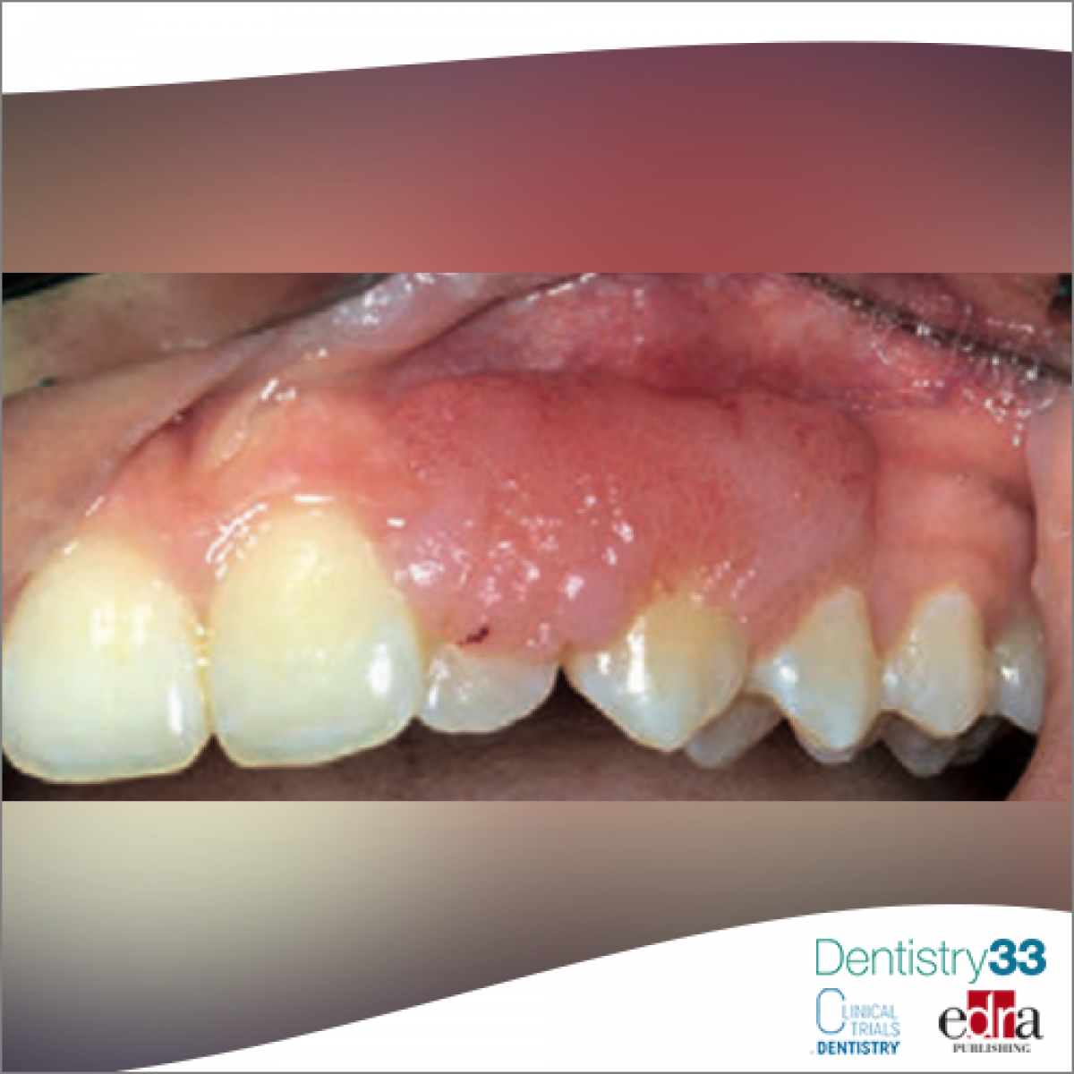
The lesion extended at the level of the vestibular gingiva from the distal margin of element 2.1 to the medial portion of element 2.4. The external surface had a finely cobbled appearance, mainly pink and with some bright red spots (fig. 1). On palpation, the consistency was similar to that of the surrounding gingiva.
With the aid of a periodontal probe it was possible to suspect that the implant base, which cannot be assessed upon inspection, was considerably smaller than the external surface. Tooth elements 1.1 to 2.5 were viable and showed no mobility. On physical examination, no other lesions were found in the oral mucosa and perioral skin.
The pediatrician, to exclude a peripheral manifestation of an hematological pathology, had already requested blood tests (complete blood count, protein electrophoresis, liver and kidney function) which were found to be normal. The radiographs made by the treating dentist excluded pathological alterations at the level of the alveolar bone and the roots of the dental elements.
Among the differential diagnoses are considered squamous papilloma (frequently found in the oral mucosa, but generally smaller in size), reactive lesions that typically develop on the gum (such as pyogenic granuloma, fibrous epulis, peripheral gigantic cell granuloma and peripheral ossifying fibroma), lymphangioma and spongiotic gingivitis. The latter occurs mainly in female subjects in puberty (10-15 years) on the vestibular aspect of the maxillary gingiva in the form of exophytic lesions, well demarcated, erythematous, with a granular or slightly papillary surface. Although many features are compatible with the case in question, the presence of a narrow implant base reduces the likelihood of this diagnosis. Lymphangioma is
a lesion that originates from the lymphatic vessels and is currently considered more of an amar- toma or a developmental anomaly than a real tumor. It manifests itself in the first two years of life and, when it develops superficially at the level of the oral mucosa, it assumes a defined “fish egg” appearance, similar to that observed in the case described. However, the onset during puberty is very rare.
Since the clinical and anamnestic characteristics are strongly suggestive for a benign pathology, after obtaining informed consent according to current legislation, we proceed with the complete separation of the neoformation and the histological examination. The pathologist observes a proliferation of keratinized stratified squamous epithelium, organized in digitiform protrusions along a fibrovascular axis, compact
bile with the diagnosis of squamous papilloma. It is a pathology induced by the Human Papilloma Virus (HPV). It has been calculated that squamous papilloma represents up to 8% of all neoformations subjected to biopsy in children. It usually manifests as an exophytic, soft, generally pedunculated formation with a warty or “cauliflower” surface with superficial projections that may be pointed or blunt. It tends to grow rapidly until it reaches a maximum size of about 0.5 cm which then remains stable over time. However, as in the case described, lesions greater than 3 cm have been observed in rare circumstances. Squamous papilloma has no degenerative potential, is treated with surgical excision and generally does not recur.
 Related articles

Authors: G. Mergoni, I. Giovannacci, G. Giunta, G. Ghidini, M. Meleti, M. Manfredi, P. Vescovi
An 11-year-old patient was sent to out observation by her pediatrician for a maxillary gingival lesion that had arisen about 2 months earlier and...
Oral pathology 02 June 2026
This peer-reviewed oral pathology article summarizes clinical evidence from Oral oncology (2026). It focuses on findings that may help dental professionals evaluate treatment decisions, patient...
Oral pathology 27 May 2026
This peer-reviewed oral pathology article summarizes clinical evidence from BMC oral health (2024). It focuses on findings that may help dental professionals evaluate treatment decisions, patient...
Oral pathology 26 May 2026
This peer-reviewed oral pathology article summarizes clinical evidence from Oral oncology (2026). It focuses on findings that may help dental professionals evaluate treatment decisions, patient...
Oral pathology 18 May 2026

This peer-reviewed oral pathology article summarizes clinical evidence from Oral oncology (2026). It focuses on findings that may help dental professionals evaluate treatment decisions, patient...
Read more

New initiative invites dentists to experience DEXIS’ most advanced AI yet, built on scale, speed, and clinical trust.
News 05 June 2026

(Nasdaq: ALGN), a leading global medical device company that designs, manufactures, and sells the Invisalign® System of clear aligners, iTero™ intraoral scanners, and exocad™ CAD/C
News 05 June 2026

(Nasdaq: HSIC), the world’s largest provider of health care solutions to office-based dental and medical practitioners, today announced that its Board of Directors has elected Will

As the University of Colorado School of Dental Medicine celebrates the graduating DDS Class of 2025, we are proud to recognize the students and faculty members whose exceptional de
Oral surgery 05 June 2026
This peer-reviewed oral surgery article summarizes clinical evidence from International journal of oral and maxillofacial surgery (2026). It focuses on findings that may help dental professionals...


Copyright © 2026 - All Rights Reserved
ISSN 2767-1178