

Facial swelling: a clinical case
Co-authors: F. Scotti, M. Mandaglio, S. Decani, E. Baruzzi, L. Moneghini
Giovanni Lodi

An 85-year-old patient comes to our attention complaining of the presence of swelling of the right half-left, present for some time unspecified and gradually increased.
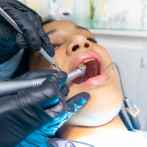
The patient's medical history is positive for: hypertension, in therapy with valsartan; polyposis of the colon; previous right pleural effusion, currently in therapy with furosemide; osteoarthritis; anemia; atrial fluid, in oral anticoagulant therapy with warfarin. At the intraoral objective examination there is a sexual neoformation, with a diminished consistency, reddish-purple color, partially ulcerated on the surface, easy to bleed, of about 6x4 cm, in correspondence with the gingiva of the IV quadrant, with extension from the buccal fornix to the oral floor (fig. 1). Furthermore, abundant plaque and tartar deposits are observed. The orthopantomography of the dental arches does not show evident signs of alteration of the hard tissues underlying the lesion.
DIAGNOSIS AND TREATMENT
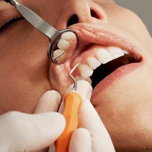
On the basis of the clinical features highlighted during the objective examination, the lesion was placed in differential diagnosis with malignant mesenchymal neoplasms, the exophytic variant of the oral cartonoma, the melanoma and, in view of the poor oral hygiene and the abundant accumulation of tartar deposits, with lesions of a reactive / irritative type, such as the pyogenic granuloma. After obtaining written informed consent from the patient and prior evaluation of the INR of the day (2.02), perilesional infiltration of local anesthetic with vasoconstrictor and execution of incisorial biopsy sampling - using a blade method cold ”- of the antero-vestibular portion of the lesion. At the end of the intervention, the haemostasis is obtained by compressing the bloody site with gauze soaked in tranexamic acid. The collected material is oriented on paper, immersed in a fixative liquid and sent to fellow pathologists for microscopic analysis of the sample (fig. 2a, 2b, 2c).
The histopathological analysis returned the outcome of malignant mesenchymal proliferation, widely ulcerated with the formation of granulation tissue and intense acute inflammation, with morphology and immunophenotype consistent with malignant fibrous histiocytoma.
Malignant fibrous histiocytoma is a soft tissue sarcoma first identified by Ozzello in 1963 and subsequently described in detail by O'Brien and Stout in 1964. It represents about 20-30% of all sarcomas of soft tissue and is considered the most frequent of these in the adult. The incidence rate is approximately 1-2 cases per 100,000 patients per year, peaking between the fifth and seventh decade of life and slight predilection for the male sex. The lower limbs more frequently follow, followed by the upper limbs and the retro peritoneal portions; the head-neck region is only affected in 4-10% of cases, although it remains one of the most frequent sarcomas at this level.
In the head-neck district, the most commonly involved sites are the nasal cavities and the paranasal sinuses, followed by the maxillary bones. Much more rarely, it occurs at the level of other structures such as oral mucosa, gums, alveolar ridges and hard palate. The aetiology is still little known. Clinically, the malignant fibrous histiocytoma appears as a deep, slow-growing mass, almost always asymptomatic, while the rapidly growing variants, much rarer, tend to be accompanied by pain symptoms. The intraoral localizations of this neoplasm enter into differential diagnosis with the pyogenic granuloma, the giant cell granuloma and the Kaposi's sarcoma. The survival rate is closely related to the presence or absence of distant metastases. About 5% of patients have metastases at the time of diagnosis, mainly in the lung. The preferred treatment is surgical excision with large resection margins; since the presence of local metastasites is not common, in most cases it is not necessary to resort to laterocervical lymphoadenectomy. The use of radiotherapy and chemotherapy is still controversial, and these treatments are reserved for cases that cannot be surgically attacked or when non-infiltrated surgical resection margins cannot be obtained.

Fig 1: Intraoral aspect: broad red-violet neoformation, diminished in consistency and partially ulcerated, in correspondence with the gingiva of the IV quadrant

Fig 2a: Hematoxylin-eosin staining of the sections of the sample taken at increasing magnifications

Fig 2b: Hematoxylin-eosin staining of the sections of the sample taken at increasing magnifications.

Fig 2c: Immunohistochemical staining for the CD163 antigen (monocytic / macrophage series), which highlights a uniformly and intensely positive cellular immunocytochemical profile for CD163
 Related articles
Oral pathology 19 June 2026
This peer-reviewed oral pathology article summarizes clinical evidence from BMC oral health (2024). It focuses on findings that may help dental professionals evaluate treatment decisions, patient...
Oral pathology 15 June 2026
This peer-reviewed oral pathology article summarizes clinical evidence from Oral oncology (2026). It focuses on findings that may help dental professionals evaluate treatment decisions, patient...
Oral pathology 10 June 2026

This peer-reviewed oral pathology article summarizes clinical evidence from BMC oral health (2024). It focuses on findings that may help dental professionals evaluate treatment decisions, patient...
Oral pathology 09 June 2026

This peer-reviewed oral pathology article summarizes clinical evidence from Oral oncology (2026). It focuses on findings that may help dental professionals evaluate treatment decisions, patient...
Oral pathology 02 June 2026
This peer-reviewed oral pathology article summarizes clinical evidence from Oral oncology (2026). It focuses on findings that may help dental professionals evaluate treatment decisions, patient...
Read more
Products 24 June 2026

News 24 June 2026

The program celebrates graduates joining the nationwide Aspen Dental network in a landscape of growing demand for oral healthcare professionals.
News 24 June 2026

Mitsui Chemicals recently announced its intention to acquire Ultradent Products Inc., a global leader in cosmetic, preventive, and restorative dentistry.
Editorials 24 June 2026
Personal trauma leads to dental hygiene career for new Texas A&M College of Dentistry honor grad

Endodontics 24 June 2026

This peer-reviewed endodontics article summarizes clinical evidence from BMC oral health (2026). It focuses on findings that may help dental professionals evaluate treatment decisions, patient...


Copyright © 2026 - All Rights Reserved
ISSN 2767-1178