

Prevalence of sinusitis of odontogenic origin and associated factors
Lara Figini
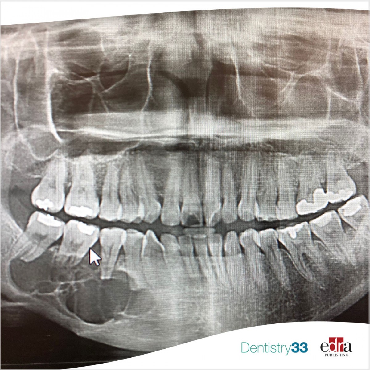
The maxillary sinuses are pneumatic cavities located inside the jaw bones, between the orbital and nasal cavities. They are covered by a thin membrane called Schneiderian, composed of a Pseudostratified ciliated columnar epithelium which constitutes an important defense mechanism of the upper airways, as well as having the function of producing mucus, filtering, heating and purifying the inspired air.
Inflammation of this membrane can be caused by several factors, including respiratory tract infections, inhalation of irritants and infectious processes extending to the maxillary sinuses. The maxillary sinuses may extend towards the alveolar process of the maxillary bones, showing a very close anatomical relationship with the apices of the maxillary teeth, especially molars and second premolars. Several literature studies have reported that the evolution of an odontogenic dental infection can lead to inflammatory changes in the Schneiderian membrane, even without its perforation. Therefore, the pathological effects of odontogenic infections could naturally be considered as etiological factors of sinusitis.
Materials and methods
In a systematic review, published in the Journal of Endodontics in February 2023, the authors determined the aggregate prevalence of odontogenic maxillary sinusitis (MSOO) and tested the associations between different odontogenic conditions and MSOO. The researchers conducted literature searches in six electronic databases and the gray literature on Aug. 25, 2022. Observational studies reporting the prevalence of MSOO and associated conditions in adults were selected by two independent reviewers.
The team excluded studies that did not use computed tomography (CT) for diagnosis. The methodological quality of the studies was assessed using the Joanna Briggs Institute Critical Appraisal Checklist for cross-sectional studies. Data were analyzed by meta-analysis for proportion and association. The certainty of the evidence was assessed using the GRADE approach.
Results
Researchers included 38 studies in the qualitative analysis and 31 in the meta-analysis. Only 12 studies (31.6%) met all elements of the methodological quality checklist. Overall, the studies reported measures of prevalence per maxillary sinus or patient. The pooled prevalence of MSOO was found to be 51% per maxillary sinus. Apical lesions, periodontitis, moderate and severe bone loss were significantly associated with MSOO. However, the certainty of the evidence for associations was very low.
Conclusions
From the data of this study, the researchers concluded that the pooled prevalence of MSOO at CT evaluation is 51% per maxillary sinus and 50% per patient. Therefore, half of maxillary sinusitis can be of odontogenic origin. Apical lesions, periodontitis and moderate and severe bone loss are significantly associated with MSOO.
Clinical implications
The associations emerging from this systematic review are based on low evidence certainty, so caution should be exercised in extrapolating these findings into clinical practice. Future studies should be conducted with greater methodological rigor. Careful clinical evaluation and historical medical and dental investigation of these patients, as well as precise evaluation of CT images, are necessary to broaden the field of knowledge and provide more accurate reports.
Filipe Colombo Vitali, Pablo Silveira Santos, Carla Massignan, Lucianne Cople Maia, Cleonice da Silveira Teixeira. "Global Prevalence of Maxillary Sinusitis of Odontogenic Origin and Associated Factors: A Systematic Review and Meta-Analysis." Journal of Endodontics. 2023 Apr;49(4):369-381.e11. doi: 10.1016/j.joen.2023.01.010. Epub 2023 Feb 6.
 Related articles
Endodontics 17 February 2026

Treatment options for endodontic failure include nonsurgical or surgical endodontic retreatment, intentional replantation, and extraction with or without replacement of the tooth.
Digital Dentistry 05 December 2025
Artifact-resistant superimposition of digital dental models and cone-beam computed tomography images
Combining the maxillofacial cone-beam computed tomography (CBCT) model with its corresponding digital dental model enables an integrated 3-dimensional (3D) representation of skeletal structures,...
Periodontology 10 September 2025

To update the findings of a systematic review from the year 2016 on the evidence for the accuracy and potential benefits of cone beam computed tomography (CBCT) in periodontal diagnostics.

Orthodontics 02 April 2025
CBCT in orthodontics: assessment of treatment outcomes and indications for its use

Since its introduction into dentistry in 1998, CBCT has become increasingly utilized for orthodontic diagnosis, treatment planning and research.
Read more

Barsoum explains the purpose of the educational events and the role 3D digital workflows can play in dentistry.

Roll-out supports parent education, provider communication, and coordinated specialty care at scale

Over six years, Align has contributed more than $2M to Operation Smile’s student programs to send the next generation of global health advocates to the world stage.
Editorials 23 July 2026
From California to Canada: CU Anschutz School of Dental Medicine at ADEA, GRC and IADR 2026
The CU Anschutz School of Dental Medicine will showcase a wide breadth of educational innovation, faculty development, clinical training and research at three major gatherings this
Oral surgery 23 July 2026

This peer-reviewed oral surgery article summarizes clinical evidence from International journal of oral and maxillofacial surgery (2026). It focuses on findings that may help dental professionals...


Copyright © 2026 - All Rights Reserved
ISSN 2767-1178