


Peri-implant tissue conditions at implants treated with Sub-periosteal Peri-implant Augmented Layer technique.
Lara Figini

The placement of an implant where there is reduced bone thickness often results in peri-implant bone dehiscence or fenestration. Even in the presence of intact but thin buccal cortical bone, implant placement can lead to vertical bone loss with exposure of the coronal part of the implant. Recently Trombelli, Severi, Pramstraller, Farina and coll. (2018) proposed a simplified bone augmentation procedure, called Subperiosteal Peri-implant Bone Thickening Technique (SPAL), based on the use of the periosteum as a membrane / barrier and a graft as a "device" to create space for bone augmentation concomitant with implant placement. In the study we are going to summarize, the effectiveness of this technique to correct peri-implant bone dehiscence and / or to increase the thickness of the peri-implant bone if necessary.
Materials and methods
In this Italian retrospective study, published on Clinical Oral Implants Research 31-2020, the authors presented an innovative implant surgical technique where there was little or insufficient bone thickness (less than 2 mm) and evaluated the conditions of the short-term peri-implant tissue by comparing the results obtained in patients in whom the SPAL technique (Sub-periosteal Peri-implant Augmented Layer or sub-periostal peri-implant bone thickening technique) was performed compared with the results obtained in patients with adequate peri-implant vestibular bone thickness (≥2 mm) and treated with traditional method.
Prior to implant placement, all patients had undergone active therapy for treating carious lesions and periodontal diseases. All surgical procedures were performed by two experienced periodontologists (L.T. and M.P.). Patients were given 2 g amoxicillin + clavulanic acid (Augmentin, GlaxoSmithKline) one hour before the surgery. Local anesthesia was achieved using articaine with 1: 100,000 adrenaline administered by local infiltration.
Two groups compared:
- Group 1 or experimental SPAL (comprising the SPAL dehiscence subgroup + SPAL thin bone thickness subgroup) comprising patients in whom there was a dehiscence defect or a thin buccal peri-implant bone thickness (PBBP) corrected with the SPAL technique performed simultaneously with the positioning of the 'plant.
- Group 2 or control group including patients treated in the traditional way as having a residual buccal peri-implant bone thickness (PBBP) ≥2 mm at the time of implant insertion.
The number of peri-implant sites positive for bleeding on probing (BoP) at 6 months after prosthetic loading was evaluated as a primary outcome. As secondary outcomes, the height of the keratinized mucosa (KM), the level of marginal soft tissues, the plaque index, the depth of peri-implant probing (PD), suppuration on probing were evaluated. Radiographic bone level (RBL) was also assessed.
SPAL technique:
A flap was performed and the periosteal layer was lifted from the bone and the sites for the accommodation of the implants were prepared. If buccal dehiscence was evident, or buccal bone thickness was too thin (less than 2mm), a deproteinized mineral bovine xenograft was performed at the same time (Bio-Oss® spongy granules, particle size 0.25-1 , 0 mm; Geistlich Pharma, AG) alone or in combination with autologous cortical bone particles and the periosteal layer was sutured to the oral flap. The buccal dehiscence was completely corrected. A free gingival graft was used to obtain adequate dimensions of peri-implant keratinized mucosa. Clinical and radiographic checks were conducted 6 months after delivery of the prosthesis.
Surgical procedures - control group:
A full thickness buccal and lingual / palatal flap was raised to expose the bony crest. The implant site was prepared according to the manufacturer's instructions and implants were placed and since there was a PBBP thickness ≥2 mm in these patients, no bone augmentation procedures were performed.
Postoperative procedures in all patients:
Patients were instructed not to wear removable prostheses to avoid compression on the surgical site for at least 4 weeks and not to use while chewing or brushing the treated area for about 2 weeks. Rinses with chlorhexidine 0.12% (Curasept ADS Regenerating Treatment®; Curaden Healthcare) for 3 weeks. The sutures were removed 2 weeks after surgery. The prosthetic rehabilitation was started 3-4 months after the implant was placed in the control group and at least 4 weeks after the implant was reopened in the SPAL group.
Results
Thirty-four patients (11 in the SPAL dehiscence group, 11 in the SPAL thin thickness group, and 12 in the control group) were included. In each SPAL subgroup 10 patients (90.9%) showed a peri-implant tissue thickness ≥2 mm in the most coronal portion of the implant. The prevalence (number) of BoP positive sites was 2, 1, and 0 in the SPAL dehiscence, SPAL thin bone thickness, and control groups, respectively. RBL amounted to 0.3 mm in the SPAL dehiscence subgroup, 0.2 mm in the thin thickness SPAL subgroup and 0 mm in the control group.
Conclusions
From the data of this study, which must be confirmed in other similar studies, it can be concluded that after 6 months of prosthetic loading, patients treated with the SPAL technique show limited inflammation of the peri-implant mucosa in association with superficial PD and adequate KM. In implants that received the SPAL technique, however, the interproximal radiographic bone level (RBL) was found to be more apical than its ideal position.
Clinical implications
Since favorable results have been observed in the short term using the SPAL technique, it is possible to hope for good results even in the long term from this technique. However, further long-term studies are needed to confirm.
For additional information: Peri-implant tissue conditions at implants treated with Sub-periosteal Peri-implant Augmented Layer technique: A retrospective case series
 Related articles
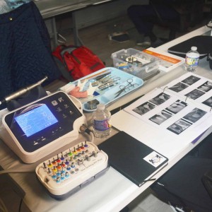
New York Implant Institute now offers live online participation for its hands-on implant training courses, allowing registered doctors to participate remotely via Zoom.
News 02 October 2026
New York Implant Institute Plans Live Patient Surgery Courses
Institute offers a variety of hands-on implant training through its dental implant CE courses and live surgery courses.
Products 23 September 2026
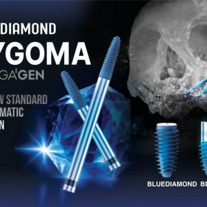
New zygomatic implant system brings MegaGen’s BLUEDIAMOND engineering, XPEED® surface technology, and flexible prosthetic solutions to complex full-arch rehabilitation.
News 16 September 2026
Products 07 September 2026
New York Implant Institute Plans Live Patient Surgery Courses
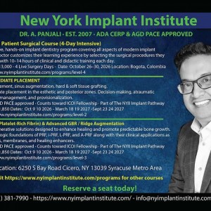
Institute offers a variety of hands-on implant training through its dental implant CE courses and live surgery courses.
Read more

Jen Kim Field, chief sustainability officer at Henry Schein and president of the Henry Schein Cares Foundation, answers questions for Dentistry Today about the group’s achievements
News 12 October 2026
The education-first program offered learning for the entire dental practice, with practical insights spanning the full patient journey and focused on improving workflows, collabora
News 12 October 2026
AAO and Stand for the Silent Unite Communities This Month to Shine a Light on Bullying Prevention
Editorials 12 October 2026
Expanded coverage: Texas A&M’s Plemons named 2026 Texas Dentist of the Year
Sometimes a small act of generosity can make a tremendous impression, especially on a 7-year-old.
Endodontics 12 October 2026
This peer-reviewed endodontics article summarizes clinical evidence from Journal of dentistry (2026). It focuses on findings that may help dental professionals evaluate treatment decisions, patient...


Copyright © 2026 - All Rights Reserved
ISSN 2767-1178