

Evaluation of the risk of perforation of the mandibular incisor canal after implantology
Lara Figini
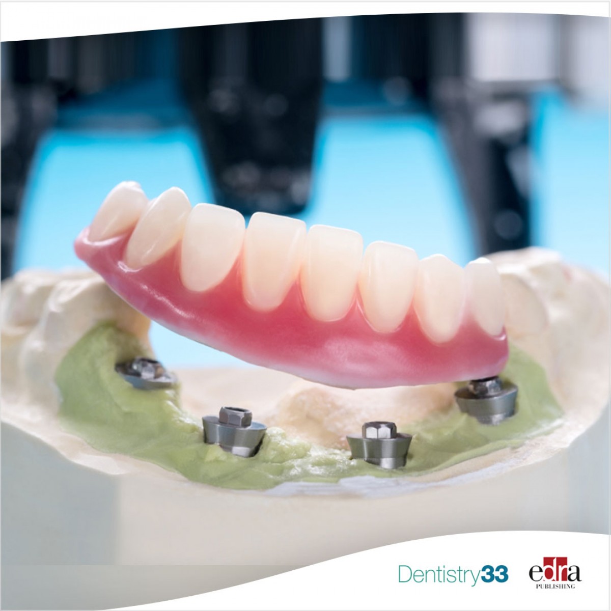
Overdentures supported by two implants, which are generally placed in the canine region, have been widely used in recent years in patients with severe atrophy of the jaw. With this method, high patient satisfaction and a high implant success rate can be achieved at a low cost.
When performing surgical procedures in the mandibular region, the inter-foraminal region is considered a safe area. But recent studies have reported various risks, ranging from minor complications to life-threatening conditions following procedures conducted in this region.
For example, the presence of the mandibular incisor canal (MIC) in the anterior mandibular region requires particular attention in implant surgery, during autogenous bone harvesting, genioplasty and fracture surgery.
However, in cases where the presence of the MIC is not paid attention to, various post-operative complications may occur such as bleeding, edema, sensory disturbances, failure of implant osseointegration and pulp sensitivity.
Due to the rapid spread of implantology, the number of procedures performed in the anterior mandible region is increasing, which in turn increases the importance of detecting vasculature, MIC and lingual concavities in this region.
Materials and methods
In a study published in May 2023 in Oral Surgery Oral Medicine Oral Pathology Oral Radiology, the authors evaluated the risk of perforation of the mandibular incisor canal (MIC) using cone beam computed tomography (CBCT) images caused by the placement of implants in the edentulous anterior mandibular region.
Some 1,200 dental implants were placed with virtually 150 suitable CBCT scans. CBCT images of patients between 2012 and 2022 were taken from the CBCT Archive at the Faculty of Dentistry of Katip Celebi University in Izmir (Turkey).
Maxillofacial radiology department images of 150 patients meeting the inclusion criteria were scanned and the data obtained were included in the study. Four implants with the following dimensions were selected: 4x8, 4x10, 4x12, and 4x14 mm.
The position of the implants was determined by an experienced surgeon (IT) by measuring on the orthopantomography (OPG) reconstructed from CBCT images, approximately at least 2 mm from the mandibular base.
The implants identified in the OPG were placed at least 1 mm away from the buccal and lingual cortices in cross section and half bucco-lingually of the crest in axial section. MIC perforation was determined when the implant perforated the canal.
The relationship of different implant sizes with the incidence of MIC mandibular incisor canal perforation and the relationship between ridge height and perforation was evaluated.
Results
A total of 1200 virtual implant applications were performed in 150 patients. MIC was identified in 87% of cases. Perforation with 12- and 14-mm implants was significantly higher than with 8- and 10-mm implants (p < 0.05).
Perforation was found to be statistically significantly greater in alveolar ridge heights ≤20 mm than that obtained with ridge heights >20 mm (p < 0.05).
Conclusions
From the data of this study, it can be concluded that high perforation rates occur in 12- and 14-mm implants and with ridge heights ≤20 mm during implant surgery in the anterior mandibular region edentulous.
For more information: "Risk Assessment of Mandibular Incisive Canal Perforation During Dental Implant Surgery in the Anterior Edentulous Mandible: A Virtual Implant Placement Study Using Cone-Beam Computed Tomography."
 Related articles
Implantology 03 February 2026
Bone Structure, Metabolism, and Physiology Its Impact on Dental Implantology
When placing implants in the mandible or maxilla, it is important for clinicians to understand the process of bone remodeling, the different types of bone, and how these factors can affect the...
Endodontics 02 February 2026
Implantology 20 January 2026
Smoking as a risk factor for dental implants and implant-related surgery

Cigarette smoking is known to adversely affect wound healing, and thus may jeopardize the success of dental implantation and implant-related oral surgery.
Products 27 November 2025
Neocis Unveils Next-Generation AI-Powered Robotic System for Dental Implants

Neocis, the pioneer behind the first and only U.S. FDA-cleared robotic system for dental implant surgery, today announced the launch and FDA approval of its next-generation robotic platform: Yomi...
Read more
Recently unveiled software features improved user experience designed to bring clarity, efficiency, and confidence to daily orthodontic treatment.
News 22 July 2026

The Tellie Awards honor the innovators expanding access to care, improving patient outcomes, and advancing medical-dental integration.
News 22 July 2026

Peak Dental Services, a growing doctor-led dental services organization, is redefining what it means to support dental professionals, becoming the first dental services organizatio
Editorials 22 July 2026
Taylor accepted as a Fellow of the American Dental Education Association Council of Deans

Reginald Taylor, associate professor and director of predoctoral orthodontics at Texas A&M College of Dentistry in Dallas, was recently accepted into the American Dental Education
Oral Hygiene & Prevention 22 July 2026

This peer-reviewed oral hygiene & prevention article summarizes clinical evidence from Journal of dentistry (2026). It focuses on findings that may help dental professionals evaluate treatment...


Copyright © 2026 - All Rights Reserved
ISSN 2767-1178