

Advantages of using 3D radiology compared to traditional two-dimensional radiology
Authors: Prof. Dr. Daniele Cardaropoli, Dr. Alessandro Roffredo
The increasing need for diagnostic precision in the dental field requires the execution of a detailed clinical examination often accompanied by supporting radiographic investigations.
CBCT (Cone Beam Computed Tomography) is an apparatus for three-dimensional radiological investigations that began to be developed at the beginning of the 90s and met an initial diffusion in dental practices starting from 2000. This equipment is in constant development technology and today's market offers highly evolved machines that are increasingly performing both in terms of versatility and the dose absorbed by the patient.
Before the spread of three-dimensional radiology, the preferred radiological investigation was represented by two-dimensional radiology (intraoral x-rays and orthopantomography).
Two-dimensional radiography has significant limitations and sometimes gives rise to aberrations that can lead the clinician to an incorrect or incomplete interpretation of the investigated site and consequent diagnostic and therapeutic errors.
This article will explain the advantages of three-dimensional radiology compared to two-dimensional radiology, taking, for example, some clinical cases we have treated.
However, it should be remembered that these radiographic investigations must always be carried out in full compliance with the principle of justification, legislation that limits the choice of CBCT as a first-level radiographic investigation.
HOW DOES A CBCT WORK?
Compared to CT (computed tomography), CBCT, as the name implies, is based on the emission of a truncated cone-shaped electron beam that is acquired by a flat-shaped receptor. Scanning takes place in a single rotation of 360 ° or 180 ° depending on the equipment around a fixed fulcrum. At each degree of rotation, the X-ray generator emits an impulse picked up by the sensor that rotates in a manner integrated with the source. Two-dimensional images of the analyzed anatomical structure are thus generated. The acquired images are subsequently processed by the acquisition software and joined together to obtain an accurate three-dimensional reconstruction of the analyzed structure.
WHY A CBCT AND NOT A CT?
The technology underlying CBCT allows acquiring tomographic images with an absorbed dose from 1.5 to 12 times lower than a spiral CT with limited scattering disturbance effects due to the presence of metal. CBCT is considered a "low dose" radiographic investigation as it performs only one rotation around the patient's head while spiral CT performs several rotations resulting in greater X-ray emission. For example, a classic CT examination for a maxillary implant site implies a radiation dose of about 2100 µSv while a CBCT of 10-60 µSv depending on the size of the FOV (Field Of View) determined and the resolution
CBCT allows you to select FOVs of different sizes, also customizable: this feature allows us to reduce the dose emitted and absorbed by the patient to the minimum necessary.
In compliance with the ALARA (As Low As Reasonably Achievable) principles of international radiation protection legislation, the operator must be able to master the software and have the ability to choose the correct FOV and the correct resolution based on the site and the pathology to investigate. Unfortunately, we often see the use of excessively large FOVs that expose the patient to excessive absorption of X-rays.
The MayRay Hyperion X9 pro used by us allows the operator to choose three different resolution programs for every single exam: "Quick" (designed for control exams), "Regular" (for basic diagnostics), and "Best" Quality "(for the research of anatomical details of reduced dimensions such as root fractures or secondary endodontic canals)
CBCT generates cubic and, therefore, isotropic voxels at very high resolution. Some machines, such as the Hyperion X9 pro, reach up to 68 µm in size: this allows you to appreciate details up to a minimum equal to this size. Furthermore, the isotropy of the voxels allows for anatomy reconstructions with a 1:1 ratio with the consequent possibility of measurements faithful to reality. CT instead generates parallelepiped voxels anisotropic rectangles with cuts of 1 mm.
Compared to CT, the acquisition of the "Cone Beam" is much more comfortable and quick and is performed in an upright position and not lying down and with a single rotation around the head to benefit the patient's safety.
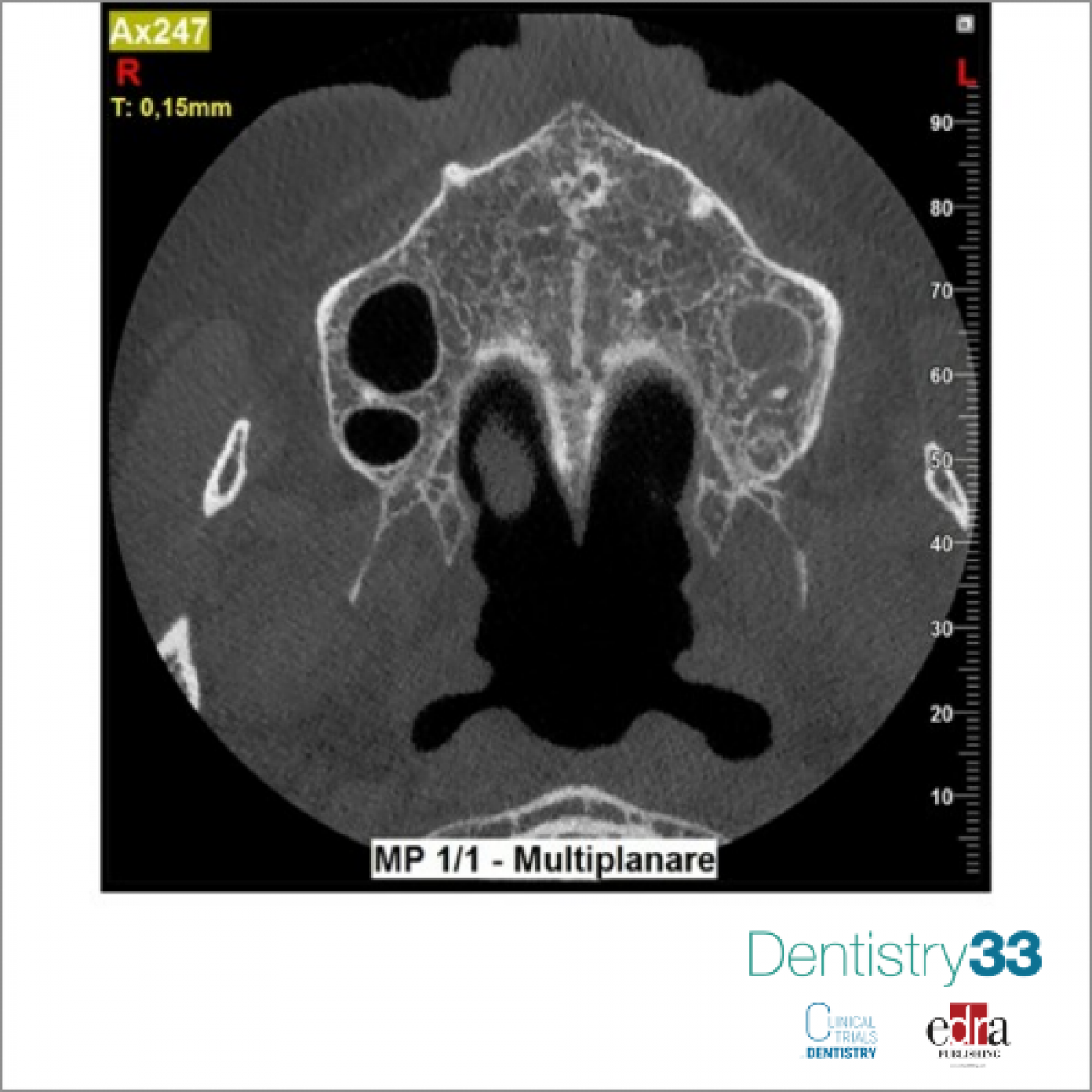
Furthermore, CBCT allows having different views of the same image, i.e., frontal, sagittal, coronal, and oblique sections.
The scanning speed, 14 "at the most for the Hyperion X9 pro, minimizes the risk of the patient moving during the execution of the exam, thus affecting the success of the investigation.
LIMITS OF 2D RADIOGRAPHIC
Two-dimensional radiography, the usefulness of which has been known since the end of the 19th century, has been the "gold standard" of dental investigations for almost a century. However, the introduction of digital radiology in the 1980s marked a further evolutionary step, bringing significant benefits both in terms of the dose absorbed by the patient and in terms of the times and methods of development of the radiographs (no need for plate development fixing liquids).
The analysis of a two-dimensional X-ray intrinsically presents the risk of making diagnostic errors since it is the projection of a three-dimensional structure on a two-dimensional plane. The superimposition of the dental, dentoalveolar, and adjacent bone anatomical structures inevitably generates distortions and/or concealments of possible pathological situations. The geometric distortions of the structures are also possible due to deformations of the receptor or following its malpositioning (incorrect or distorted projection of the dental elements), thus generating the difficulty in obtaining certain measurements.
The human factor also intervenes in two-dimensional radiology: an expert eye can notice details that a less experienced eye can hardly recognize. An "interpretation" of the radiographic investigation is therefore required, a potentially risky situation due to the incorrect information it can give.
These limits have been overcome by three-dimensional radiology that allows accurate and detailed visualization of the anatomical structures and pathologies, thus allowing the clinician to decide the best treatment plan to apply to the patient's care.
FIELDS OF APPLICATION 3D RADIOLOGY
CBCT has an almost exclusively dental field of application, however, it also allows the investigation of anatomical structures such as maxillary sinuses and ear canals that are of interest to the maxillofacial of otolaryngology.
In the dental field, the fields of application are different:
• All doubtful situations in which two-dimensional radiology is insufficient and does not allow to have a precise diagnosis of the site or pathology investigated
• Implant surgery: the 1:1 ratio of the images generated by CBCT and the isotropy of its voxels allow precise measurements corresponding to reality, a necessary condition for implant surgery to avoid the lesion of sensitive anatomical structures during the insertion of the implants adjacent to the recipient site and to evaluate the amount of residual bone in the edentulous areas.
• Guided surgery: CBCT is essential for the preliminary planning of the implant position in cases of mask-guided surgery
• Extractive surgery, especially of the eighth octaves included, in complex anatomies that could lead to injury to the inferior alveolar nerve or other anatomical structures during surgical maneuvers
• Endodontics: for the evaluation of extremely complex dental anatomies in case of root canal retreatments, the evaluation of endodontic complications such as perforations, the evaluation of root or bone resorption, the search for root fractures
• Trauma: in situations where traditional radiology does not give diagnostic certainty
• Reconstructive surgery, for example, evaluates the anatomy of the maxillary sinuses in the case of sinus lift operations.
CONCLUSIONS
Modern three-dimensional radiology allows overcoming the limits of traditional two-dimensional radiology by improving the quality of the images obtained and providing the operator with an impressive amount of information about the patient's anatomy.
The latest generation CBCTs, such as the MyRay and the Hyperion X9 pro, thanks to the technological evolution of the acquisition sensors, minimize patients' exposure to X-rays, thus allowing compliance with safety regulations and significantly reducing the related risks.
Training the operator in the use of software and in the choice of the correct FOVs and resolutions in relation to the site and the pathology to be investigated is of fundamental importance to correctly return to the biological costs / clinical benefits (ALARA principle).
 Related articles
News 08 May 2026

Maria Gutierrez in Yorba Linda, California, has added the DEXIS Orthopantomograph OP 3D EX cone beam computed tomography (CBCT) system to its practice.
Products 23 February 2026
Next Generation Trident X-View 3D CBCT Product Line Now Available Through Mid America Dental Sales

The next generation Trident X-View 3D CBCT product line is now available through Mid America Dental Sales, a master distributor of dental technology. These advanced imaging systems from Italy are...
Products 22 December 2025

AI dental company announces 510(k) clearance from the U.S. Food and Drug Administration for Overjet CBCT Assist.
Products 24 September 2025

With top-of-the-line 3D imaging technology and all the essential dental imaging programs, Planmeca’s latest offering easily meets the everyday needs of general dentistry, endodontics, and...
Periodontology 10 September 2025

To update the findings of a systematic review from the year 2016 on the evidence for the accuracy and potential benefits of cone beam computed tomography (CBCT) in periodontal diagnostics.
Read more
Products 26 June 2026
Coronal flaring is considered a key step in efficient root canal preparation, especially for difficult-to-access canals.
News 26 June 2026
The Association for Dental Safety (ADS) proudly announced the recipients of the 2026 Leadership Awards during its Annual Conference on May 27 in Salt Lake City, Utah.
News 26 June 2026

Recognition highlights the company’s doctor-led culture and continued investment in team member growth and engagement
Editorials 26 June 2026
From California to Canada: CU Anschutz School of Dental Medicine at ADEA, GRC and IADR 2026
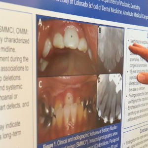
The CU Anschutz School of Dental Medicine will showcase a wide breadth of educational innovation, faculty development, clinical training and research at three major gatherings this
Oral surgery 26 June 2026

This peer-reviewed oral surgery article summarizes clinical evidence from International journal of oral and maxillofacial surgery (2026). It focuses on findings that may help dental professionals...


Copyright © 2026 - All Rights Reserved
ISSN 2767-1178