


Multidisciplinary Management of a Traumatized Tooth in the Aesthetic Zone
This case report illustrates the successful interdisciplinary management of a complex dental trauma involving tooth No.
Preserving natural teeth is a fundamental goal in dentistry, reflecting clinical and patient-centered priorities. Advances in periodontal and prosthodontic co-therapy have made it possible to save teeth that would traditionally be extracted.
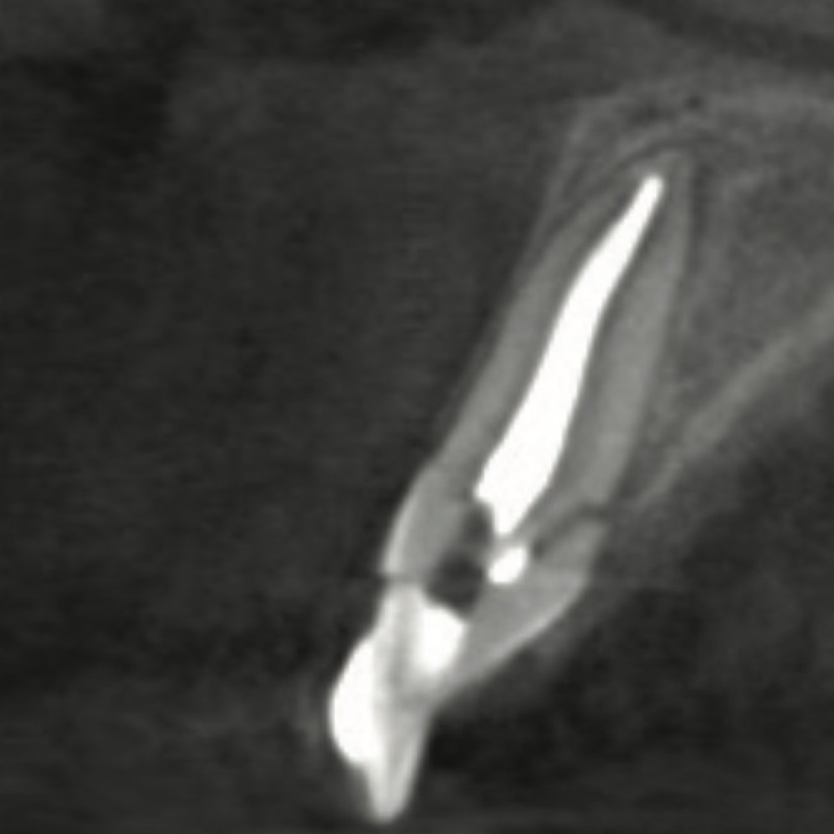
A 13-year-old boy sustained facial trauma resulting in an oblique fracture of tooth No. 8. The patient was referred to our periodontal office by his orthodontist for crown lengthening surgery to facilitate orthodontic forced eruption. A CBCT scan (CS 8200 3D Neo [Carestream Dental]) revealed that the fracture extended to the alveolar crest (Figures 1 and 2). The proposed treatment plan was to move the fractured tooth coronally by an additional 2 mm before performing CLS. This ensured a minimum distance of 3 mm between the crown margin and the bone to avoid biological width invasion (BWI). Alternatives such as extraction and implant placement were deemed less favorable due to the patient’s age and the potential for growth-related complications.
Orthodontic forced eruption (OFE) was initiated to elevate the fractured tooth coronally (Figure 3), facilitating CLS while minimizing the risk of damage to the periodontium around adjacent teeth. OFE is critical to avoid the need for an ostectomy around adjacent teeth or compromising the biological width during restoration. The purpose of OFE was to create sufficient coronal tooth structure for proper crown seating and bonding of orthodontic brackets on tooth No. 8 (Figures 4 and 5). Additionally, controlled eruption allows for better outcomes when performing subsequent restorative procedures, as it maintains the natural gingival contours and minimizes aesthetic complications. This method has been shown to improve long-term periodontal stability and restorative outcomes. 1
The proposed orthodontic treatment was to slowly erupt No. 8 utilizing partial fixed orthodontic appliances (0.018-in Innovation self-ligating brackets [Specialty Appliances]), which were placed on the anterior teeth and anchor tubes placed on Nos. 3 and 14 (0.022-inch) (Figure 4). To slowly super-erupt No. 8, a 0.016- × 0.022-in braided wire (Quad Cat) was placed with 1.0-mm step-down bends.
Topical anesthetic gel (ADS) was used prior to using one carpule of 2% Lidocaine with Epi 1 x 100,00 (Cook-Waite Lidocaine). Following removal of the orthodontic bracket and archwire, a scalpel blade (No. 12 Bard-Parker [Aspen Surgical]) was used for intrasulcular incision followed by the Goldman-Fox No. 7 knife for flap elevation. The CLS was performed using burs from the Brasseler Kit (Brasseler USA). The Oschenbein No 2 was used to avoid injuring roots. The fractured portion of tooth No. 8 was removed, revealing minimal remaining coronal tooth structure (Figure 6). The CLS exposed adequate tooth structure for restorative procedures while preserving periodontal health (Figure 7). 2 The procedure was followed by core buildup around the post and temporization with Integrity (BIS-GMA from RESIMENT) material to facilitate orthodontic bracket bonding. 3 Notably, CLS was performed with precise consideration of the biological width, ensuring that the soft tissue was not encroached upon, which could otherwise lead to inflammation and bone resorption (Figures 8 and 9). The surgical area was closed using the 4.0 Silk Suture (Salvin Dental). An ice pack (Jack Frost from the Net32) and written post-op instruction were delivered before dismissing the patient.
Tapered chamfer diamond burs (850.FG.014 [Komet USA]) were used to finish the preparation of tooth No. 8 followed by polishing paste (Diamond Polish [Ultradent Products]). In addition, a bite registration impression (Blu-Mousse [Parkell]) was taken.
Final impressions (Impregum [Solventum]) were taken after 6 weeks of healing. A ceramic crown was fabricated and cemented using RelyX Luting Cement (Solventum) (Figures 10 to 12). The patient’s orthodontic treatment resumed, with the restored tooth integrated into the arch. This phase was crucial as proper healing and crown placement ensured both functional and aesthetic success. Studies have highlighted the importance of using durable restorative materials and proper cementation techniques in young patients to achieve long-term stability and prevent secondary complications. 4
Natural teeth offer numerous advantages over prosthetic replacements: 5
Recent research highlights the long-term success of periodontally treated teeth, even in cases of severe compromise:
The success of this case underscores the importance of interdisciplinary care. Each specialist contributed uniquely: 8
This collaborative approach aligns with evidence-based practices, benefiting all parties involved.
The decision to preserve a fractured tooth vs opting for extraction and subsequent implant placement is influenced significantly by the patient’s age. In pediatric and adolescent patients, as seen in the case of a 13-year-old referred for CLS on tooth No. 8, age plays a critical role in treatment planning. Younger patients often have ongoing development in their craniofacial structures, including the alveolar bone. This continued growth can affect implants’ success and stability due to potential bone volume changes over time. Studies have shown that maintaining the natural tooth is often more advantageous in younger patients because it avoids the risks associated with implant placement during growth periods, such as altered bone development and aesthetic concerns. 9
In cases like the one described, where the patient’s development is still active, the emphasis on maintaining the natural tooth through orthodontic techniques (eg, OFE) allows for gradual repositioning without the need for surgical augmentation. This approach minimizes the risk of complications and provides better outcomes for function and aesthetics. It is also aligned with evidence suggesting that younger patients exhibit better success rates with tooth preservation strategies compared to adult patients due to the higher regenerative potential of younger tissues.
CBCT plays a pivotal role in the diagnosis and treatment planning of complex cases involving tooth fractures and the need for subsequent surgical intervention. 10 CBCT imaging allows clinicians to visualize the fracture in 3D, which is critical for accurate assessment of the extent and location of the damage. For the case of fractured tooth No. 8, the CBCT scan revealed an oblique fracture extending to the alveolar crest at the palatal aspect. This detailed imaging provided a comprehensive view of the fracture pattern, root morphology, and proximity to surrounding anatomical structures, such as the maxillary sinus, mandibular canal, and adjacent teeth.
By utilizing CBCT, clinicians can plan surgical interventions with greater precision. It enables the surgeon to plan for adequate bone reduction (ostectomy) if needed, assess the potential need for grafting materials, and avoid unnecessary removal of bone around adjacent teeth. The 3D visualization provided by CBCT also aids in planning the OFE more accurately, ensuring that the movement of the tooth does not compromise the adjacent periodontal structures. 10
In summary, the use of CBCT in the diagnosis and treatment planning of complex dental cases, especially in pediatric patients, is critical. The ability to visualize and plan precisely allows for more conservative and effective treatment, preserving the natural tooth whenever possible.
The management of aesthetic concerns in the anterior teeth requires a delicate balance between maintaining natural tooth structure and achieving optimal aesthetics. This case underscores the importance of a multidisciplinary approach in dental care, where both restorative and periodontal expertise are combined to achieve the best possible outcome for the patient.
The preservation of tooth No. 8 in this adolescent patient highlights the potential of modern periodontal-prosthodontic co-therapy to save teeth traditionally deemed unsalvageable. The use of OFE, CLS, and careful restorative techniques demonstrates the clinical and biological advantages of retaining natural teeth. As dental care continues to evolve, clinicians must prioritize minimally invasive, patient-centered approaches that align with the principles of preserving natural dentition whenever feasible.
Integrating patient age considerations with advanced diagnostic tools like CBCT significantly impacts the decision-making process in complex periodontal-restorative cases. By utilizing these methods, dental professionals can provide more personalized treatment plans that prioritize the preservation of natural teeth, especially in younger patients.
Dr. Ahmad Soolari is a Diplomate of the American Board of Periodontology. He has a certificate in periodontics from the Eastman Institute for Oral Health and a MS degree from the University of Rochester in Rochester, NY. He is a former clinical associate professor at the University of Maryland Dental School in Baltimore. Dr. Soolari operates a specialty practice in the Silver Spring, Gaithersburg, and Potomac area of Montgomery County, Md. He can be reached at asoolari@gmail.com.
Dr. Ehsan Soolari has graduated from the University of Maryland School of Dentistry. He focuses on preventive and personalized care in addition to pain management and cosmetic dentistry at Soolari Dentistry. He serves the communities of Silver Spring, Md. He is the author of multiple articles in medical and dental journals. He can be reached at esoolari@gmail.com.
Disclosure: The authors have no financial interest in any companies mentioned in this article and received no compensation for writing this article.
Source: https://us02web.zoom.us/webinar/register/WN_pTvYxBGcQ_ak65szMIj5Ww#/registration
 Related articles
.jpg)
Talking, laughing, chewing, and smiling are daily facial movements everyone makes with little to no thought, however for individuals with temporomandibular disorders (TMDs), those simple movements...

UNC Adams School of Dentistry’s Jane A. Weintraub, DDS, MPH, the R. Gary Rozier and Chester W. Douglass Distinguished Professor, first became interested in the science of aging when she took a...
News 08 September 2023

The IU Board of Trustees approved the construction of the clinic, which is supported through a $2 million grant from the Delta Dental Foundation. Construction is slated to begin in mid-2024.
Endodontics 21 April 2023

By Norberto Maccagno, editorial director, Odontoiatria33
Professor Giovanni Olivi, president of the International Academy of Innovative Dentistry, talks about the new academy and its goals and the organization’s first Congress, which will be held in Rome...
McEnhill’s father was a dentist and she said that she spent part of her teenage years working at his dental practice. “I learned very quickly how to work with patients in an extremely busy...
Read more
Europe 27 September 2026 - 30 November -0001
Digital Dentistry Society Global Congress 2026: Event Preview and Professional Highlights

The Digital Dentistry Society Global Congress 2026 will take place from October 22, 2026 to October 24, 2026 in TBD, Rotating Europe, offering dental professionals a focused environment for...
Editorials 27 September 2026
Dentistry33 survey highlights replacement care, deep caries and evolving restorative workflows
Reader survey finds direct composites remain dominant, secondary caries is the leading reason for restoration replacement, and clinicians want more training in pulp preservation
Reader survey finds direct composites remain dominant, secondary caries is the leading reason for restoration replacement, and clinicians want more training in pulp preservation
Edra Professional Books 26 September 2026
Manual of oral surgery: A Practical Reference for Dental Professionals
Manual of oral surgery is an Edra professional dentistry reference focused on clinical practice, education and treatment planning.
Products 25 September 2026
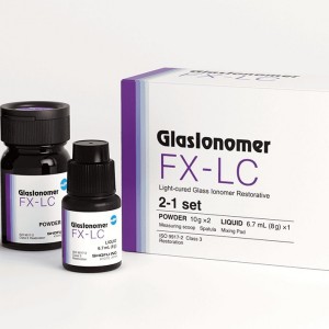
Shofu Dental’s line of dental restorative materials now includes FX-LC, a next-generation, light-cure, resin-reinforced glass ionomer formulated to deliver high strength, efficient 4
News 25 September 2026

The scholarships are part of an ongoing workforce solutions strategy to help address the need for skilled dental hygienists


Copyright © 2026 - All Rights Reserved
ISSN 2767-1178