

Indirect restoration of 1.6 and 1.7: a clinical case
Allegra Comba

38-year-old male patient with negative medical history, who comes for a visit many years after the last dental check-up. He reports difficulty in removing food residues in the interproximal areas, in correspondence with some fractured amalgam restorations. However, it does not report pain / thermal sensitivity on any of the dental elements.
Physical and radiographic examination of the dental surfaces shows the presence of secondary carious lesions affecting the following dental elements: 1.7, 1.6, 1.5, 1.4. (Fig. 1-2)
The cold test performed on vital dental elements is positive and highlights the absence of irreversible pulp pathologies. Diagnosis Occlusal-distal carious lesion on 1.4 and mesio-occlusal-distal carious lesions on 1.5, 1.6 and 1.7.
Proposed treatment plan
Periodontal probing, scaling and root planing to remove subgingival tartar deposits;
Oral hygiene education and motivation;
Revaluation survey before proceeding with conservative therapies;
Execution of direct composite restorations on 1.4 and 1.5 and indirect restorations on 1.6 and 1.7.
The execution of direct restorations was explained at the following link: https://www.dentistry33.com/clinical-cases/restorative-dentistry/180/direct-restoration-of-1-4-and-1-5-a-clinical-case.html
STEP-BY-STEP CLINICAL PROCEDURES FOR INDIRECT RESTORATION OF 1.6 and 1.7
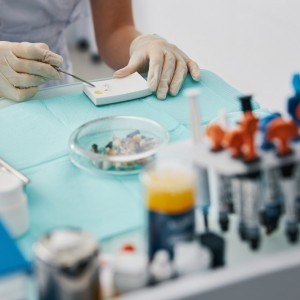
1)Removal of carious lesions on 1.6 and 1.7
Isolation of the operating field with the rubber dam fixed at the level of element 1.7 with a previously sandblasted 27N hook (Fig. 1). Subsequently, removal of the carious lesions on 1.6 and 1.7.
For the removal of the amalgams, a multi-blade tungsten drill mounted on a red ring contra-angle was used initially, under abundant jet of water and suction, followed by a cutter cylindrical diamond bur and a ceramic rosette for the removal of infected tissue at the dentin substrate level.
The cavity margins were finished with red and yellow ring diamond cutters.
The cervical steps of the cavities were also terminated with red and yellow grit diamond cutters mounted on the EVA handpiece and abrasive diamond strips (Fig.2).
2)Build-up and impression
After the application of an etch-and-rinse 3 steps adhesive system for 1.6 and a self-etch 2 steps adhesive system with pre-etching of the enamel, two composite build-ups were carried out, which were subsequently prepared and finished with diamond burs with decreasing grain size. The cervical steps of the cavities have also been finished with red and yellow grain diamond burs mounted on the EVA handpiece and diamond abrasive strips (Fig. 3). Once the rubber dam was removed, the residual thicknesses for the future indirect restoration were assessed and the impression of the entire upper arch with PVS and the antagonist alginate arch were taken. In order to avoid problems related to the sensitivity of the prepared vital elements, a temporary restoration was used.
3)Inlay composite cementation
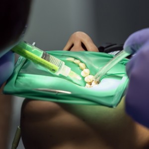
At the next appointment, we proceeded by removing the temporary restorations and isolating the operating field with the rubber dam. The prepared elements were cleaned with prophylaxis brushes and glycine conveyed with air flow and then the adhesive procedures were carried out on 1.6 and 1.7: etched with 37% orthophosphoric acid, primer application, silane (on the composite build up) and bonding, the latter uncured. At the same time, the adhesive preparation of the composite inlay took place: sandblasting with aluminum oxide (50micron) to remove any debris, application of the silane and bonding, also in this case without proceeding with its polymerization (Fig. 4). A layer of heated nanohybrid composite, color A3, was placed on the bottom of the build-up of 1.7 and the composite product was housed on the preparation with the help of an adhesive plastic stick. In order to correctly position the product, constant pressure was performed with the help of a ball burnisher and a dedicated ultrasound tip (Fig. 5). The same procedures were also carried out for cementing the inlay on 1.6.
Once the polymerization phases of about 2 minutes per dental element were completed, a further polymerization of 20 '' per side was performed after applying a layer of glycerine gel to obtain a further conversion of the oxygen-inhibited layer. To finish the surfaces of the restored elements, diamond cutters with low particle size were used, followed by paper disks with different degrees of abrasiveness, by rubber pads and brushes. The interproximal area was further refined with low-grain diamond burs on the EVA handpiece and with low-abrasive metal strips, in order to maintain a correct contact point and emergence profile.
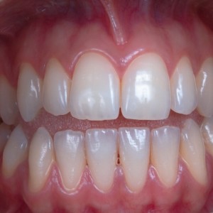
The reconstructed elements were checked one week after the last appointment to check for any problems during the function (Fig. 6a-b).

Fig 1: pre and post isolation with dental dam

Fig 2: cavity preparation

Fig 3: build-up 1.6 and 1.7

Fig 4: cementation

Fig 5: inlay 1.6 and 1.7

Fig. 6a: control after 1 week

Fig 6b: control after 1 week
 Related articles
Restorative dentistry 17 April 2026
Patient Perceptions of New Robotic Technologies in Clinical Restorative Dentistry
Patient perception research has failed to focus on burgeoning technology within the dental field.
Prosthodontics 16 April 2026
The use of orthodontics before fixed prosthodontics in restorative dentistry
For a variety of reasons, orthodontic intervention is often overlooked as a viable modality to correct occlusal, axial, rotational, and space discrepancies before undertaking fixed prosthetic...
Digital Dentistry 16 February 2026
An original procedure for the ovoid pontic technique in ortho-restorative cases

In the multidisciplinary treatments, especially in case of missing teeth in aesthetic area, the compliance of patient during retention time post orthodontic therapy is evidently very high, even among...
Restorative dentistry 29 January 2026
Current trends in restorative dentistry in the UK: a Delphi approach
The purpose of the study was to obtain insight into current trends in restorative dentistry in the UK by means of a Delphi technique.
Read more
Products 23 June 2026
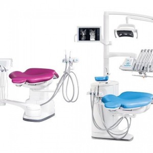
Planmeca introduces two dental units designed to support modern clinical workflows through different concepts and use cases.
Dental practice transaction can come with a variety of obstacles in the most normal of circumstances.
The American Society for Dental Aesthetics invites you to attend the 2026 Annual International Conference on Dental Aesthetics Excellence at the Conrad Nashville, October 21-24, 20
Editorials 23 June 2026
Periodontology 23 June 2026
This peer-reviewed periodontology article summarizes clinical evidence from Oral health & preventive dentistry (2026). It focuses on findings that may help dental professionals evaluate treatment...


Copyright © 2026 - All Rights Reserved
ISSN 2767-1178