

Effect of different incisal preparation designs on stress distribution within the ceramic veneer-tooth system
Lorenzo Breschi
Introduction
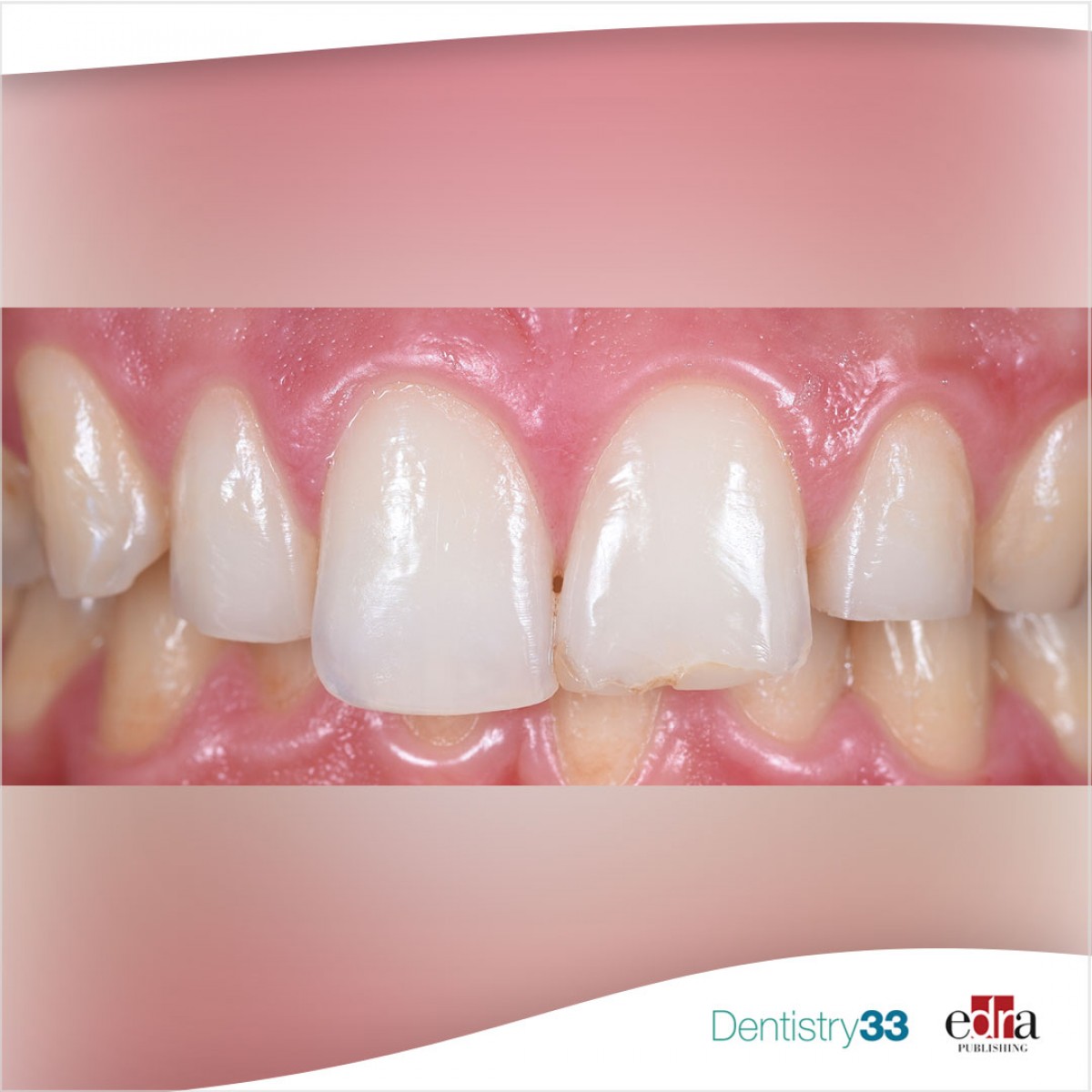
Ceramic veneers are widely used in clinical practice due to their aesthetic nature and clinical performance. Although they are strong and durable, showing high survival rates, ceramic chipping and fracture are the most common reasons for failure of the veneer restorations.
Incisal preparation design is regarded as one of the predisposing factors for failure of these restorations. Although the incisal preparation design for ceramic veneers has been widely discussed, there is no consensus among in vivo studies due to the heterogeneity of clinical results.
In this regard, Yin Chai et al. have conducted an in vitro study to evaluate the stress distribution within the ceramic veneer-tooth system with butt joint (BJ) and feathered edge (FE) incisal preparation designs at two loading angulations – 0° and 20° – using a photoelastic stress analysis technique.
Materials and methods
Six photoelastic models representing maxillary left central incisors were fabricated using an epoxy resin material and were divided into six groups according to BJ and FE preparation configurations and loading angulations.
Lithium disilicate ceramic veneers (IPS e.max CAD, Ivoclar Vivadent) were bonded to the BJ and FE photoelastic models with a dual-cure resin cement (IPS Variolink Esthetic, Ivoclar Vivadent) according to parameters and guidelines recommended by the manufacturer.
Each group was loaded at the incisal edge using an Instron Universal Testing Machine at a crosshead speed of 0.25 mm/min up to a maximum of 100 N. Images of the photoelastic model were exported from the webcam when the load was at 0, 10, 50 and 100 N, respectively. The isochromatic fringe patterns, representing location and intensity of stress concentrations, were recorded.
Results
At 0° loading angulation, the BJ photoelastic model showed better stress distribution compared with the control and FE photoelastic models. Minimal stress was visible at the cement line and at the incisal edge below the area of load application. As the load increased, stress concentration was confined to the cement layer and distributed toward the cervical area of the tooth.
At 20° loading angulation, the FE photoelastic model showed significantly higher stress development at the interface between the ceramic veneer and the photoelastic model close to the incisal edge compared with the control and BJ photoelastic models. Higher stress concentration was observed as the load increased. These stresses spread from both the palatal concavity and the cement line toward the cervical area of the tooth.
Conclusion
The authors concluded that the incisal preparation designs investigated (butt joint and feathered edge) affect stress distribution within the ceramic veneer-tooth system. The results demonstrated that BJ photoelastic model showed a more uniform distribution and much lower stress concentration compared with FE photoelastic models under both 0° and 20° loading angulations. Moreover, different loading angulations may result in different distribution of stress within the photoelastic models.
Chai SY, Bennani V, Aarts JM, Lyons K, Das R. “Stress distribution within the ceramic veneer-tooth system with butt joint and feathered edge incisal preparation designs.” J Esthet Restor Dent. 2021 Apr;33(3):496-502. doi: 10.1111/jerd.12655. Epub 2020 Sep 16. PMID: 32937021.
 Read more
Products 26 June 2026
Coronal flaring is considered a key step in efficient root canal preparation, especially for difficult-to-access canals.
News 26 June 2026
The Association for Dental Safety (ADS) proudly announced the recipients of the 2026 Leadership Awards during its Annual Conference on May 27 in Salt Lake City, Utah.
News 26 June 2026

Recognition highlights the company’s doctor-led culture and continued investment in team member growth and engagement
Editorials 26 June 2026
From California to Canada: CU Anschutz School of Dental Medicine at ADEA, GRC and IADR 2026
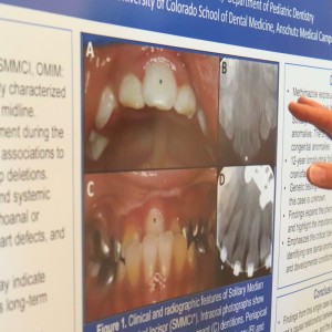
The CU Anschutz School of Dental Medicine will showcase a wide breadth of educational innovation, faculty development, clinical training and research at three major gatherings this
Oral surgery 26 June 2026
This peer-reviewed oral surgery article summarizes clinical evidence from International journal of oral and maxillofacial surgery (2026). It focuses on findings that may help dental professionals...


Copyright © 2026 - All Rights Reserved
ISSN 2767-1178