

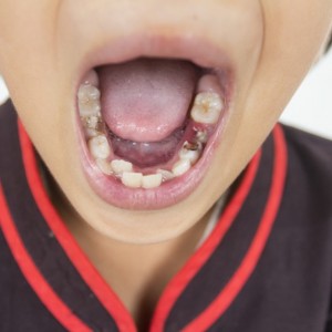
Multiple radiolucencies in a 12-year-old boy
A healthy 12-year-old boy (The American Society of Anesthesiologists Physical Status Classification I) was initially referred by a general dentist to the University of Illinois Chicago oral and maxillofacial surgery clinic for evaluation of an incidentally found asymptomatic lesion associated with the apex of tooth no. 8. His medical record included a distant history of sports-related trauma to the anterior maxilla and a family history of jaw lesions in his father and sister.
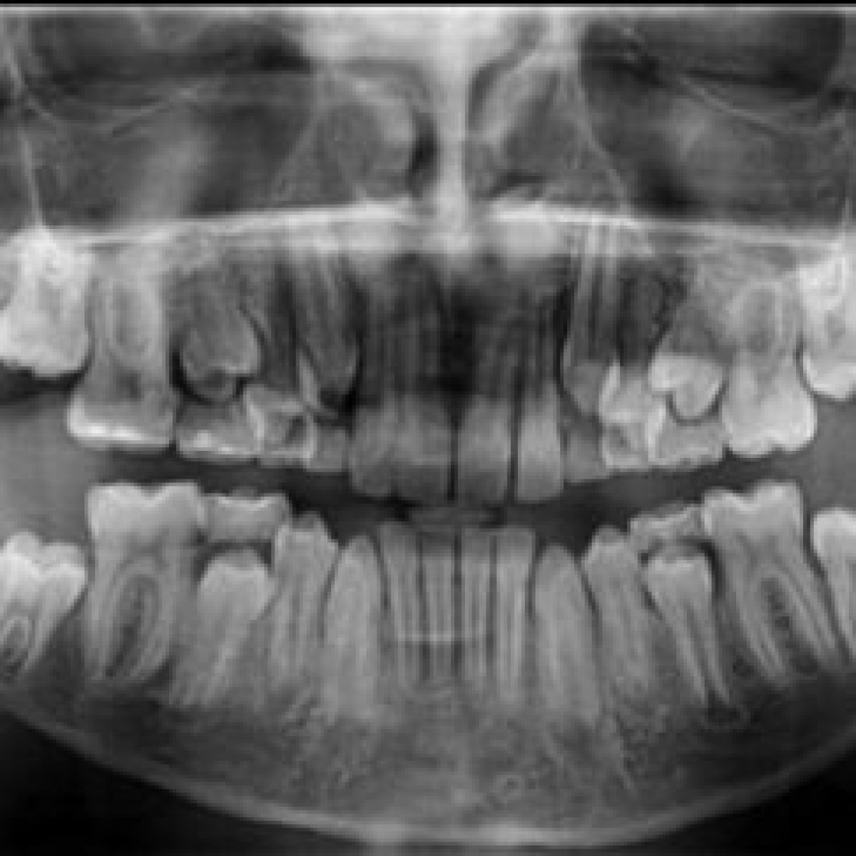
He was also previously evaluated by a dermatologist for nonpainful, nonpruritic bumps on his extremities and desquamation of his feet. At the initial consultation at the oral and maxilloficial surgery clinic, radiographic imaging revealed enlarged follicles of teeth nos. 17 and 32 and a periapical radiolucency associated with tooth no. 8 without clinical signs of asymmetry (Figure 1). The father of the patient declined a plan to perform a biopsy of the apical lesion associated with tooth no. 8.
The patient was then lost to follow-up but returned a year and a half later with asymmetry in the lower facial one-third, with the right mandible larger than the left. On intraoral examination, the region of tooth no. 8 was not tender to palpation, without fluctuance, and the pulpal and periapical testing results for tooth no. 8 were normal. Asymmetric firm bony expansion of the posterior mandible was appreciated bilaterally, with more expansion being apparent on the right.
A cone-beam computed tomographic scan revealed large homogeneous, well-demarcated, pericoronal radiolucent lesions with hyperostotic borders associated with and displacing the follicles of teeth nos. 17 and 32 distally and inferiorly to the inferior border of the mandible (Figure 2A). A third similar-appearing lesion was visualized in the anterior maxilla apical to tooth no. 8 (Figure 2B). No cortical breakthrough was seen radiographically. Incisional biopsies were performed for the 3 lesions, revealing thick cheesy exudate surrounded by a friable cystic lining.
Authors: Osman Khan, DDS, Andrew Bertagna, DMD, MD, Douglas Damm, DDS, Ashleigh Weyh, MD, DMD, MPH, Nicholas Callahan, MPH, DMD, MD, FACS
Source: https://jada.ada.org/
 Related articles
Pediatric dentistry 29 April 2026
Exploration and innovative application of digital technology in pediatric dentistry
Pediatric dentistry treats patients aged 0-18 years, encompassing the characteristics of general dentistry.
Pediatric dentistry 14 April 2026
Comparison between dexmedetomidine and esketamine in pediatric dentistry surgery
Dexmedetomidine (D) and esketamine (K) are used for the sedation of pediatric dental surgery. This study was designed to compare the effect of intranasal D and K in producing moderate sedation for...
Existing studies on adverse events (AEs) in pediatric dentistry have been limited in scope.
Products 13 January 2026
Revolutionizing Pediatric Dentistry with Dr. Josh Solomon: SDI Stela & Bioclear Insights

Join pediatric dentist Dr. Josh Solomon as he discusses the cutting-edge SDI Stela self-curing composite system and the Bioclear matrix system, and how these products are transforming Class II...
Read more
USA 07 June 2026 - 30 November -0001
ICOI World Congress USA 2026: Event Preview and Professional Highlights

The ICOI World Congress USA 2026 will take place from August 13, 2026 to August 15, 2026 in TBD, USA, offering dental professionals a focused environment for continuing education, clinical updates...
Edra Professional Books 06 June 2026
Understanding Dental Insurance: A Practical Reference for Dental Professionals

Understanding Dental Insurance is an Edra professional dentistry reference focused on clinical practice, education and treatment planning.
New initiative invites dentists to experience DEXIS’ most advanced AI yet, built on scale, speed, and clinical trust.
News 05 June 2026

(Nasdaq: ALGN), a leading global medical device company that designs, manufactures, and sells the Invisalign® System of clear aligners, iTero™ intraoral scanners, and exocad™ CAD/C
News 05 June 2026

(Nasdaq: HSIC), the world’s largest provider of health care solutions to office-based dental and medical practitioners, today announced that its Board of Directors has elected Will


Copyright © 2026 - All Rights Reserved
ISSN 2767-1178