


Quality of etched enamel: bonding and development of white spot lesions
Davide Elsido
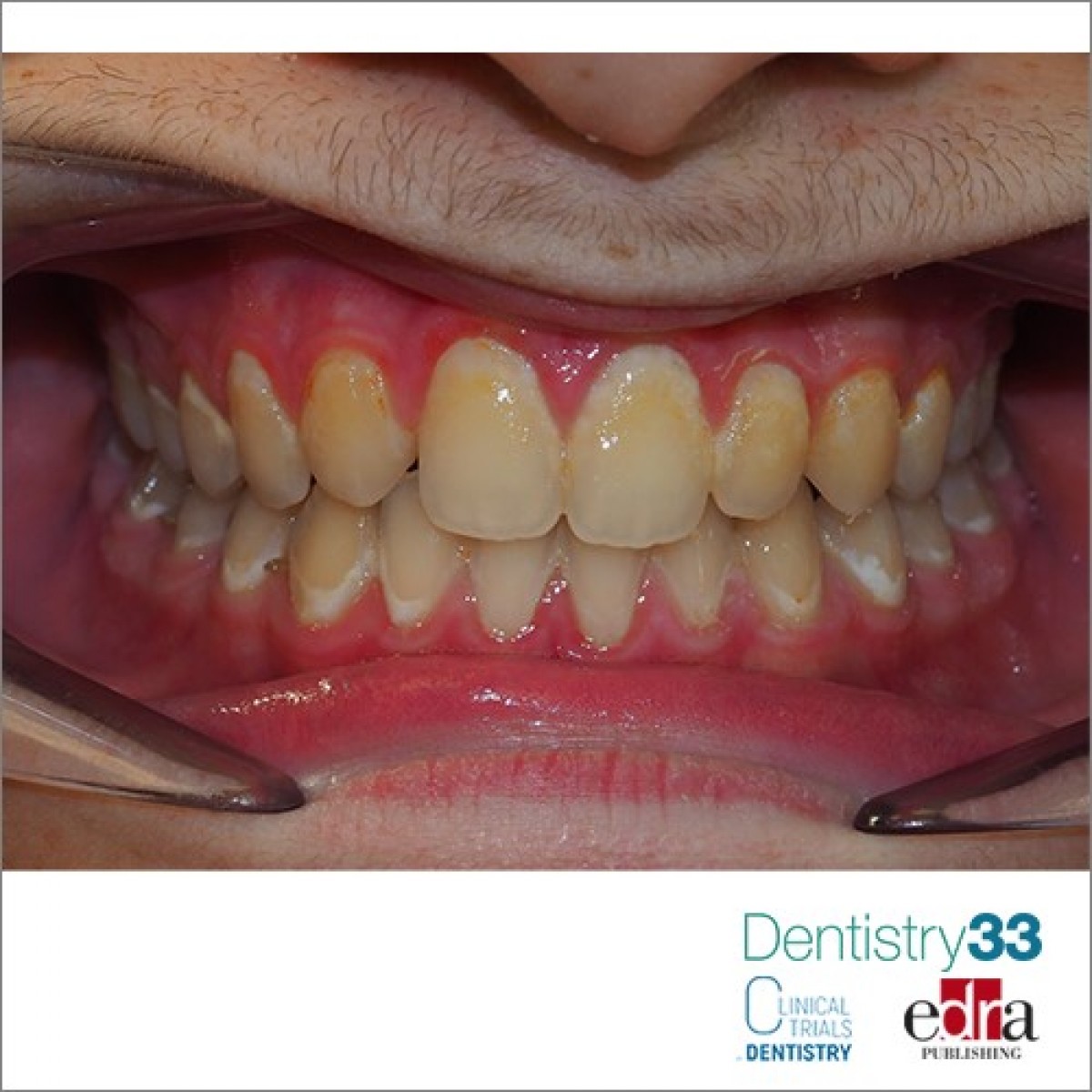
Enamel is a homogenous structure composed primarily of inorganic matter, organized into hydroxy- apatite crystal prism bundles. It has two distinct layers: an outer ‘‘prismless’’ enamel layer and an underlying prismatic layer. The prismless layer has an optic axis nearly parallel to the enamel’s surface. In contrast, prismatic enamel has distinct rod boundaries oriented perpendicular to the surface. The aprismatic enamel is believed to be due to decreased ameloblast activity during tooth development and the disappearance of Tomes’ processes during the end of amelogenesis. Etching is supposed to remove the outer aprismatic layer and expose the underlying prismatic rods. Scanning electron microscopy (SEM) images of etched buccal premolar and molar enamel have shown that the cervical region is composed primarily of prismless enamel; the incisal and middle thirds of etched premolar enamel exhibit distinct prism-end, honey- comb-type structures. Etch patterns evaluated using SEM images of silicone impressions indicate that the etch quality of the anterior teeth is superior to the quality of the posterior teeth, around this topic an article was published in the September issue of The Angle Orthodontist.
The buccal right and left halves of 27 extracted human teeth were randomly allocated to scanning electron microscopy (SEM) or micro–computed tomography (lCT) for evaluation. The buccal surfaces were pumiced, etched with 37% phosphoric acid gel etchant for 15 seconds, rinsed, and air dried. Each tooth was divided into three regions (incisal, middle, and cervical) and viewed after etching at magnification with SEM. The lCT scans were taken before and after etching to calculate apparent and material mineral densities.
Results: SEM showed greater aprismatic enamel and poorer etch quality (significantly less percentage enamel) for the posterior than anterior teeth and for the cervical region than for the incisal and middle regions of all teeth. Although there were no density differences prior to etching, lCT demonstrated that etching increased material density significantly more for the anterior than posterior teeth. Prior to etching, the enamel in the cervical regions was significantly less dense than the enamel in the middle or incisal regions. Etching significantly increased the material density of all three regions, which decreased initial regional differences. After etching, the apparent density of the cervical region remained significantly lower than the densities of the other two regions.
The study sentences that, based on SEM and lCT, there is greater aprismatic enamel and inferior etch quality in the cervical regions of all tooth types and is clinically significant in explaining the failure of sealant retention and the propensity for white spot lesions.
Clinically, the results imply that the cervical region of teeth may require a different etching protocol than the middle and incisal regions. The cervical regions, particularly of the posterior teeth, may require a longer etching time and/or a more concentrated etch to remove more of the aprismatic enamel.
 Related articles
Orthodontics 03 May 2023
Impact of clear aligners on oral health, oral microbiome during orthodontic treatment

The present work highlighted further links between clear aligners and changes in oral health and the oral microbiome and provided plaque control methods for clear aligner trays.
Restorative dentistry 16 March 2021
Evaluation of the remineralizing capacity of amorphous calcium phosphate on white spot lesions

Authors:Marco Calabrese Lorenzo Graiff Alberto Fioretti Matteo Fistarol
Dental caries is a disease that causes the destruction of the tooth due to the metabolic bacterial processes that take place within the oral biofilm. The term "caries" identifies the actual...
Read more
Products 21 September 2026
The OsteoGen™ Composite graft line uses the same biomimetic, bioactive crystal technology to harness the body’s ability to heal itself.
News 21 September 2026

The Aspen Group (TAG), a leading healthcare support organization headquartered in Chicago’s West Loop, today announced two key additions to its marketing leadership team: David Chr
News 21 September 2026

BlancOne® Click+ introduces a simple 10-minute professional teeth-whitening experience designed to integrate into existing hygiene appointments, helping dental practices create new
Editorials 21 September 2026

The new school year is in full swing at Texas A&M College of Dentistry, and that includes a host of faculty promotions and new hires and important happenings.
Endodontics 21 September 2026
This peer-reviewed endodontics article summarizes clinical evidence from Journal of dentistry (2026). It focuses on findings that may help dental professionals evaluate treatment decisions, patient...



Copyright © 2026 - All Rights Reserved
ISSN 2767-1178