

Clinical and imaging features of unilateral impacted maxillary central incisors
Davide Elsido
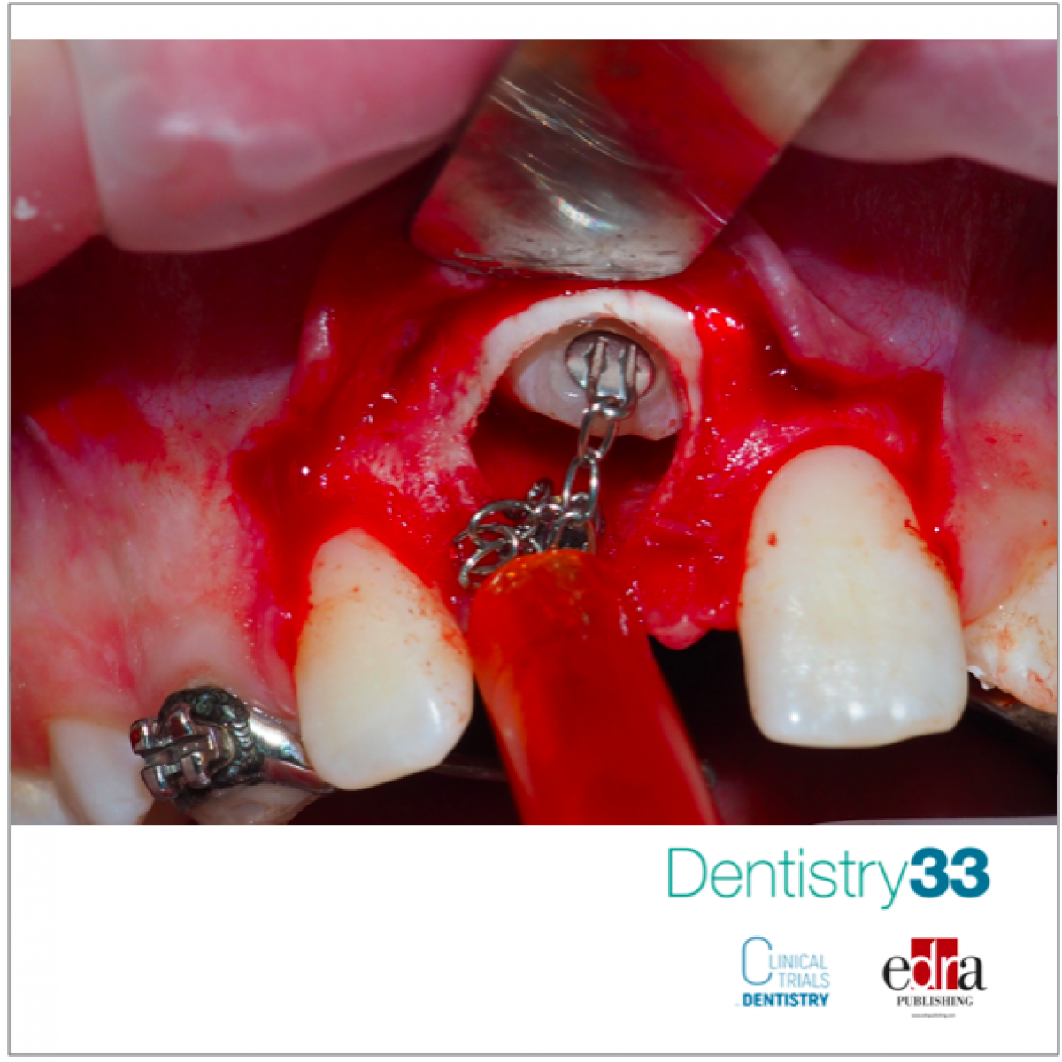
Maxillary central incisors erupt relatively early during the permanent dentition process, and they are located in a relatively forward position. The shape, size, coloration, position, and symmetry of maxillary central incisors can significantly impact facial esthetics. The prevalence of maxillary central incisor impaction is 0.06%-0.24%, which is lower compared with those of impacted maxillary canines. Although the prevalence of the impaction of the maxillary central incisor is low, its occurrence can be impactful. Delayed maxillary central incisor eruption can profoundly alter speech, chewing, and facial appearance in affected patients. The 3-dimensional (3D) position and root morphology of the impacted maxillary incisor influence the treatment difficulty for orthodontists. However, no study has provided statistical data analyzing the relationship between 3D position and the root morphology of impacted maxillary central incisors. Radiographic analyses of impacted teeth are essential to guide proper diagnosis and treatment planning. However, 2-dimensional (2D) images may not provide sufficiently detailed information because of the potential distortion or poor positioning. Cone-beam computed tomography (CBCT) offering 3D images has been recently introduced and used for the diagnosis and analysis of impacted teeth, as this approach provides multiple planes of view that can allow for more accurate analyses. In addition, these CBCT data can be used to generate 3D reconstructions using software.
A cross-sectional study was published in the February 2022 issue of the AJODO to analyze the clinical features and 3D spatial distribution of unilateral impacted maxillary central incisors and identify the factors associated with root morphology to provide a reference for patient diagnosis and treatment efforts.
Results
This patient cohort included 52 male patients and 42 female patients. Thirty-three incisors (35.11%) with dilacerated roots, 17 incisors (18.09%) with retained deciduous teeth, 15 incisors (15.96%) with supernumerary teeth, and 15 incisors (15.96%) with a history of trauma were identified in the study. Of the 94 impacted incisors, the most common were labially impacted (n 5 65; 69.15%), followed by vertically impacted (n 5 17; 18.09%) and palatally impacted (n 5 12; 12.77%).
The etiology of tooth impaction can be either primary or secondary, with the former typically being associated with abnormal germ position and ankylosis and the latter being related to trauma or obstruction of the eruption pathway. Trauma may also result in the occurrence of dilaceration. This research found that 15 patients (15.96%) had a history of trauma from 3 to 6 years of age and indicated that the influence of trauma history on the root morphology was not statistically significant, which may be attributed to the small sample size. They identified 17 patients (18.09%) with retained deciduous teeth and 15 patients (15.96%) with supernumerary teeth. These can be preventatively removed at early time points as a means of reducing the risk of maxillary incisor impaction. Prior research indicated that children with primary teeth are more likely to suffer a dental trauma, and trauma at an early age will result in the development of a dilacerated tooth, emphasizing the importance of protecting children from such trauma.
It can be challenging for clinicians to diagnose, treat and predict patient outcomes when impacted maxillary incisors have dilacerated roots: dilaceration was significantly associated with age, initial crown height, and incisor length; in addition, maxillary incisors with dilacerated roots are at a higher risk of root resorption during orthodontic treatment.
Conclusions
Labially impacted maxillary incisors were more common than vertically or palatally impacted maxillary incisors, and labially inverted incisors were most frequently found in the sagittal plane. Dilaceration was most commonly observed in inverted incisors. In clinical practice, CBCT images play an important role in diagnosing impacted maxillary central incisors and in making appropriate treatment plans for patients.
 Read more
USA 26 July 2026 - 30 November -0001
American Academy of Periodontology Annual Meeting 2026: Event Preview and Professional Highlights

The American Academy of Periodontology Annual Meeting 2026 will take place from October 15, 2026 to October 18, 2026 in TBD, USA, offering dental professionals a focused environment for continuing...
Edra Professional Books 25 July 2026
Retreatments: A Clinical Reference for Contemporary Endodontic Practice

Retreatments is an Edra professional dentistry reference focused on clinical practice, education and treatment planning.
Products 24 July 2026

Expanded shade range supports fast, esthetic chairside zirconia restorations with 9-minute sintering when used with CEREC ® SpeedFire
News 24 July 2026

Sensei, a Carestream Dental brand and a global leader in dental practice management software and digital imaging solutions, today announced a strategic partnership with MAX Surgica
News 24 July 2026

Beach Point Capital Management LP (“Beach Point”), a global alternative investment manager with over $20 billion in assets under management, today announced the successful sale of


Copyright © 2026 - All Rights Reserved
ISSN 2767-1178