

A large radiopaque lesion in the posterior mandible: a clinical case
Authors: G. Lodi, A. Pispero, E.M. Varoni, S. Decani, D. Costa
Giovanni Lodi

A 32-year-old male patient is sent, for a right mandibular injury, by his own doctor to the Oral Pathology and Surgery Service of the Operative Unit of Odontostomatology II of ASST Santi Paolo e Carlo of Milan. The medical history is negative for any type of pathology, facial deformations are not appreciated, no symptoms are reported and mucosal lesions are not appreciated intraorally. The panoramic radiography (fig. 1) and computed tomography (fig. 2), held by the patient at the time of the first visit, show a large irregular radiopacity that dominates the element 4.8. The lower right third molar is in total bone inclusion with the middle and apical third of the roots in close contact with the lower vascular-nerve bundle. The mandibular canal is strongly compressed and deviated apically in correspondence with the retained element, passing between the two mesial roots. The radiopaque mass, which dominates the crown, extends superiorly for about 1 cm up to the coronal margin of the bone cortex and distally occupies the space between the crown of the third molar and the cortex of the mandibular canal. Around the lesion there is a thin radiolucent area that creates a clear separation from the surrounding bone tissue. The patient has no other radiographic examinations prior to the visit that can allow to appreciate over time the degree of growth of the lesion.

Figure 1: Orthopantomography

Figure 2: CT cone beam
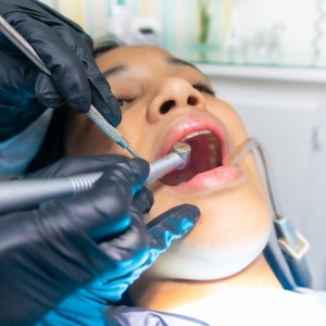
In the case described, the clinical and radiographic signs suggest a diagnosis of a complex odontoma. The mass of the lesion prevented the normal eruption of the third molar. Although the clinical suspicion is strong, it is necessary to consider the possibility of comparing with other pathologies such as osteomyelitis sclerosing, periapical dysplasia of the cement, ossifying fibroid and cementusblastoma. In agreement with the patient, it was decided to carry out an incisional biopsy (fig. 3).

Figure 3: incisional biopsy
An envelope flap with a disto-vestibular discharge incision to the crown of the element 4.7 is performed and following the detachment of a mucoperiosteal flap, a lesion is taken to send the necessary material to the anatomical pathologist .
The consistency of the lesion is hard, comparable to that of the dentine of a healthy dental element. The histological examination confirms the clinical diagnosis of a complex odontoma. This type of lesion is classified as a benign tumor of odontogenic origin. It is considered to be a dental hamartoma made up of mineralized tissues that derive from odontogenic epithelium and ectomesenchima; enamel, dentin and cement are present and well developed but follow a macroscopically disordered pattern without leading to the formation of defined dental elements.
The lesion has characteristics of benignity and has no tendency to recurrence. In the case described there are no absolute indications for the treatment of the lesion but considering the size and the relationship with the distal root surface of the element 4.7 it is decided to remove the lesion leaving the third molar in place. The extraction of wisdom tooth is not a justified intervention, the invasiveness and the risks of alteration of the nerve conduction are propensity for a conservative attitude. The prediction is that the closure by primary intention and the amount of vestibular and lingual bone cortex will allow a good wound healing and, before the completion of the remineralization phase, a natural extrusion of the tooth.
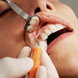
The treatment plan is discussed with the patient, who gives consent for the intervention. The flap for the removal of the lesion is similar to that performed during the incisional biopsy. With the elevation of a mucoperiosteal flap and a modest vestibular ostectomy it is possible to access the entire lesion. In consideration of the hard consistency of the lesion, separation lines are made by means of slot cutters; the fragments obtained are thus removed without having to sacrifice additional bone tissue. The sizing is carried out by means of 4/0 resorbable polyfilament. The new histological examination performed on all the lesion confirms the first diagnosis. After 6 months it is possible to appreciate in the intraoral radiography (fig. 4) how, in accordance with the forecasts formulated, the cortex has completely reformed above the included element and how it appears in position more coronal than before surgery.

Figure 4: periapical radiography after 6 months
 Related articles
Oral pathology 19 June 2026
This peer-reviewed oral pathology article summarizes clinical evidence from BMC oral health (2024). It focuses on findings that may help dental professionals evaluate treatment decisions, patient...
Oral pathology 15 June 2026
This peer-reviewed oral pathology article summarizes clinical evidence from Oral oncology (2026). It focuses on findings that may help dental professionals evaluate treatment decisions, patient...
Oral pathology 10 June 2026

This peer-reviewed oral pathology article summarizes clinical evidence from BMC oral health (2024). It focuses on findings that may help dental professionals evaluate treatment decisions, patient...
Oral pathology 09 June 2026

This peer-reviewed oral pathology article summarizes clinical evidence from Oral oncology (2026). It focuses on findings that may help dental professionals evaluate treatment decisions, patient...
Oral pathology 02 June 2026
This peer-reviewed oral pathology article summarizes clinical evidence from Oral oncology (2026). It focuses on findings that may help dental professionals evaluate treatment decisions, patient...
Read more
Products 26 June 2026
Coronal flaring is considered a key step in efficient root canal preparation, especially for difficult-to-access canals.
News 26 June 2026
The Association for Dental Safety (ADS) proudly announced the recipients of the 2026 Leadership Awards during its Annual Conference on May 27 in Salt Lake City, Utah.
News 26 June 2026

Recognition highlights the company’s doctor-led culture and continued investment in team member growth and engagement
Editorials 26 June 2026
From California to Canada: CU Anschutz School of Dental Medicine at ADEA, GRC and IADR 2026
The CU Anschutz School of Dental Medicine will showcase a wide breadth of educational innovation, faculty development, clinical training and research at three major gatherings this
Oral surgery 26 June 2026

This peer-reviewed oral surgery article summarizes clinical evidence from International journal of oral and maxillofacial surgery (2026). It focuses on findings that may help dental professionals...


Copyright © 2026 - All Rights Reserved
ISSN 2767-1178