

Lower lip mucosa neoformation
Giovanni Lodi
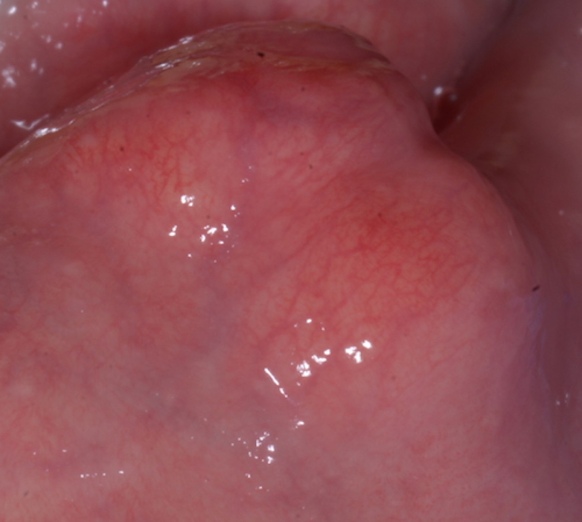
A 60-year-old patient in good general health is visited at the Oral Pathology and Surgery Service of the Operative Unit of Odontostomatology II of ASST Santi Paolo e Carlo. The medical history reveals mild arterial hypertension and type II diabetes mellitus being treated with Telmisartan 20 and Metformin 500 respectively. The non-smoker patient complains of the presence of a slow and continuous painless swelling at the mucous side of the left lower hemilab (fig.1 and fig.2). There are no skin changes in color or temperature, external palpation shows an elastic consistency in the area of the lesion, negative palpation of ipsilateral laterocervical lymph nodes.
At the intraoral level, a sessile swelling of about 2cm in diameter is observed, roundish, covered with a mucous membrane similar in appearance to the surrounding one and with a yellowish-pink color. On palpation the consistency is elastic and the lesion and the lesion is mobile below the mucous plane. No other alterations in shape or color of the intraoral mucous membranes are appreciated.
DIAGNOSIS AND CARE
The lesion on the basis of clinical data and that provided by the patient has characteristics of benignity. However, it is necessary to obtain a histological diagnosis that precisely determines the nature of the neoformation. The patient, after signing the informed consent, consents to the surgical treatment. Before proceeding with the biopsy intervention, a needle aspiration is performed at the center of the swelling through the mucous plane which does not show the presence of blood, salivary or similar purulent material. It is therefore possible to proceed with the intervention which in this case will be an excisional biopsy.
After perilesional infiltration of local anesthetic with vasoconstrictor, a mucosal incision of about 1cm is made above the lesion perpendicularly to the vermilion edge of the lip. The dissection of the tissues by blunt way through the primary incision allows to highlight a mass of frankly yellowish complexion. It is possible to easily cleave the lesion which appears frankly elastic, compact and separated from the surrounding tissues by a pseudocapsule of lining (fig. 3). At the end of the operation, the bottom of the operating field is clean and bloodless and the suturing of the incision margins takes place by means of a 5/0 absorbable polyfilament.
The diagnosis, confirmed by the histological examination, is of fibrolipoma (fig. 4). It is a benign tumor formed by a mix of fat and connective cells and is the most common histological variant of the lipoma. This pathology particularly affects limbs and trunk, when multiple sites are affected it is called multiple lipomatosis. At an intraoral level, it originates from the adipose and connective cells of the submucosa and clinically presents with a sessile or pedunculated yellowish pink swelling. The vestibular mucous membranes and the oral floor are mainly affected, following the tongue and lips. Diagnosis is simple considering the clinical aspects of color and consistency, slow growth and absence of symptoms. The differential diagnosis is with vascular lesions or from salivary tissues, due to obstructive problems of the duct or due to increases in the volume of the parenchyma. Needle spraying is a good way to distinguish these pathologies. The treatment indicated in the literature is the complete excision of the neoformation, an operation that is generally simple thanks to the ease with which it is possible to cleave the whole body of the lesion from the surrounding tissues. Relapses are very rare except for lipomas that infiltrate the muscles.

Fig1: intraoral aspect

Fig2: extraoral aspect

Fig3: lesion isolation

Fig4: histological image
 Related articles
Oral pathology 19 June 2026
This peer-reviewed oral pathology article summarizes clinical evidence from BMC oral health (2024). It focuses on findings that may help dental professionals evaluate treatment decisions, patient...
Oral pathology 15 June 2026
This peer-reviewed oral pathology article summarizes clinical evidence from Oral oncology (2026). It focuses on findings that may help dental professionals evaluate treatment decisions, patient...
Oral pathology 10 June 2026

This peer-reviewed oral pathology article summarizes clinical evidence from BMC oral health (2024). It focuses on findings that may help dental professionals evaluate treatment decisions, patient...
Oral pathology 09 June 2026

This peer-reviewed oral pathology article summarizes clinical evidence from Oral oncology (2026). It focuses on findings that may help dental professionals evaluate treatment decisions, patient...
Oral pathology 02 June 2026
This peer-reviewed oral pathology article summarizes clinical evidence from Oral oncology (2026). It focuses on findings that may help dental professionals evaluate treatment decisions, patient...
Read more
Products 26 June 2026
Coronal flaring is considered a key step in efficient root canal preparation, especially for difficult-to-access canals.
News 26 June 2026
The Association for Dental Safety (ADS) proudly announced the recipients of the 2026 Leadership Awards during its Annual Conference on May 27 in Salt Lake City, Utah.
News 26 June 2026

Recognition highlights the company’s doctor-led culture and continued investment in team member growth and engagement
Editorials 26 June 2026
From California to Canada: CU Anschutz School of Dental Medicine at ADEA, GRC and IADR 2026
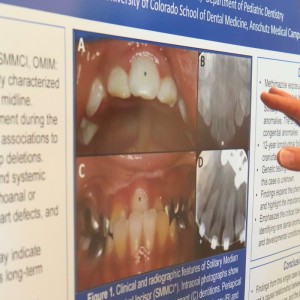
The CU Anschutz School of Dental Medicine will showcase a wide breadth of educational innovation, faculty development, clinical training and research at three major gatherings this
Oral surgery 26 June 2026

This peer-reviewed oral surgery article summarizes clinical evidence from International journal of oral and maxillofacial surgery (2026). It focuses on findings that may help dental professionals...


Copyright © 2026 - All Rights Reserved
ISSN 2767-1178