

Foreign body in the maxillary sinus
Authors: Margherita Gobbo, Giulia Ottaviani, Katia Rupel, Matteo Biasotto

A female patient was sent to the Medicine and Oral Pathology outpatient clinic of the Dental Clinic of Trieste for the evaluation of an intensely radiopaque foreign body present in the right maxillary sinus.
The patient had never experienced any symptoms. However, during a first visit performed at the Dental Clinic of Trieste, to which she had presented with a recent orthopantomography (fig.1a), the dentist had deemed it appropriate to refer it to the Medicine and Oral Pathology clinic for further information. also the intense inflammatory reaction visible radiographically.
The patient, in good health, was not taking any drugs and referred only to being allergic to tramadol.
The right maxillary arch appeared partially edentulous, with the absence of elements from 1.4 to 1.8 and the presence of an osseointegrated implant in the edentulous 1.4. During the first oral pathology visit, an intraoral radiograph was performed in site 1.4, from which no lesions were found. Clinically, the edentulous area was pink, free from continuous solutions or lesions. Given the clinical situation, age and good health of the patient, it was decided to intervene with the surgical removal of the foreign body under local anesthesia.
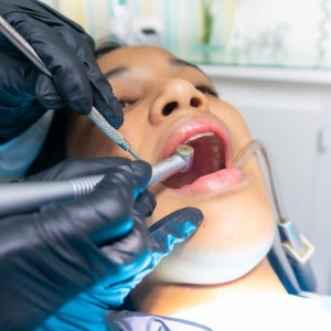
Three weeks later, the maxillary sinus was revised endoscopically. Subject to written informed consent from the patient, local anesthesia was performed and a full-thickness trapezoidal flap was prepared in area 13-17. Subsequently, a bony trap door with piezoelectric inserts was made (Figs. 2a, b), Schneider's membrane was incised and a thin and flexible fiberoptic probe was introduced to inspect the right maxillary sinus. Once the foreign body was highlighted, it was removed using a peeler and surgical tweezers (fig. 2c). The inflammatory reaction, visually polypoid in appearance, was always removed and cured endoscopically (fig. 3). Finally, the maxillary sinus was cleansed with the alternating use of sterile water and rifamycin. The bony hatch created for access to the maxillary sinus was repositioned with 5 mm mini osteosynthesis plates (fig. 2d) and, subsequently, protected with freeze-dried native collagen and with the repositioned flap. The patient was prescribed antibiotic therapy (amoxicillin 3 g / day for 6 days; N-acetylcysteine 300 mg / 3 ml to be taken every evening via aerosol). After a week, the silk sutures were removed.
The patient denied pain, swelling, or other symptoms. After 8 days, the histological examination confirmed the presence of an intense inflammatory reaction and a lesion of likely mycotic origin, also known as fungus ball. After 4 months, the picture appears stable.
Sinusitis of mycotic origin is a continuously increasing phenomenon. It is divided into two categories: invasive and non-invasive. Among the "non-invasive", there are an allergic form and the so-called fungus ball. While the first form occurs as a consequence of hypersensitivity to allergens of fungal origin mediated by the nasal sinus tract, the fungus ball type is found in non-allergic immunocompetent subjects and appears to be linked to the accumulation of debris and mycotic components in the paranasal sinuses, usually of the Aspergillus family. Despite being initially asymptomatic pathologies, the long-standing fungus ball can begin with nonspecific symptoms such as nasal congestion, purulent or blood discharge from the ipsilateral nostril, headache, facial pain or alteration of smell. The pathogenesis of these lesions remains debated. The treatment of choice remains surgery with foreign body removal and reventilation of the affected breast, with no need for adjuvant antifungal therapy.

1a

2

3
 Related articles
Oral pathology 19 June 2026
This peer-reviewed oral pathology article summarizes clinical evidence from BMC oral health (2024). It focuses on findings that may help dental professionals evaluate treatment decisions, patient...
Oral pathology 15 June 2026
This peer-reviewed oral pathology article summarizes clinical evidence from Oral oncology (2026). It focuses on findings that may help dental professionals evaluate treatment decisions, patient...
Oral pathology 10 June 2026

This peer-reviewed oral pathology article summarizes clinical evidence from BMC oral health (2024). It focuses on findings that may help dental professionals evaluate treatment decisions, patient...
Oral pathology 09 June 2026

This peer-reviewed oral pathology article summarizes clinical evidence from Oral oncology (2026). It focuses on findings that may help dental professionals evaluate treatment decisions, patient...
Oral pathology 02 June 2026
This peer-reviewed oral pathology article summarizes clinical evidence from Oral oncology (2026). It focuses on findings that may help dental professionals evaluate treatment decisions, patient...
Read more
Products 24 June 2026
News 24 June 2026

The program celebrates graduates joining the nationwide Aspen Dental network in a landscape of growing demand for oral healthcare professionals.
News 24 June 2026

Mitsui Chemicals recently announced its intention to acquire Ultradent Products Inc., a global leader in cosmetic, preventive, and restorative dentistry.
Editorials 24 June 2026
Personal trauma leads to dental hygiene career for new Texas A&M College of Dentistry honor grad

Endodontics 24 June 2026
This peer-reviewed endodontics article summarizes clinical evidence from BMC oral health (2026). It focuses on findings that may help dental professionals evaluate treatment decisions, patient...


Copyright © 2026 - All Rights Reserved
ISSN 2767-1178