

Digital Device in Postextraction Implantology: A Clinical Case Presentation
Aim
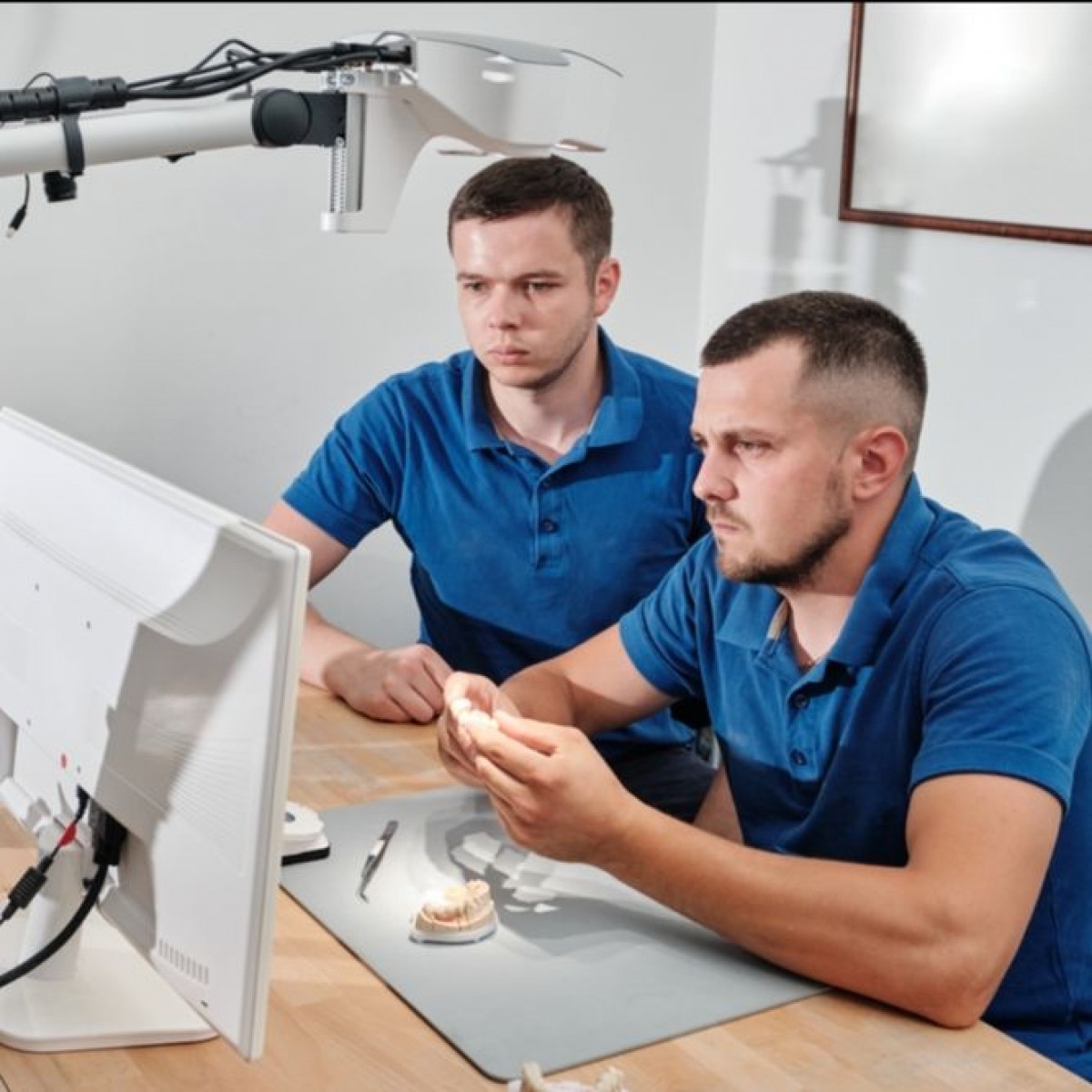
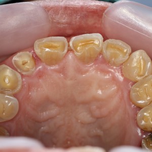
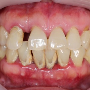
The aim of this work is to describe a case of immediate implant placement after extraction of the upper right first premolar, with the use of CAD/CAM technology, which allows an early digital impression of the implant site with an intraoral scanner (MHT 3D Progress, Verona, Italy). Case Report. A 46-year-old female was referred with a disorder caused by continuous debonding of the prosthetic crown on the upper right first premolar.
Clinically, there were no signs, and the evaluation of the periapical radiograph showed a fracture of the root, with a mesial well-defined lesion of the hard tissue of the upper right first premolar, as the radiolucent area affected the root surface of the tooth. It was decided, in accordance with the patient, that the tooth would be extracted and the implant (Primer, Edierre implant system, Genoa, Italy) with diameter of 4.2 mm and length of 13 mm would be inserted. After the insertion of the implant, it was screwed to the scan abutment, and a scan was taken using an intraoral scanner (MHT 3D Progress, Verona, Italy).
The scanned images were processed with CAD/CAM software (Exocad DentalCAD, Darmstadt, Germany) and the temporary crown was digitally drawn (Dental Knowledge, Milan, Italy) and then sent to the milling machine for production with a composite monoblock. After 4 months, when the implant was osteointegrated, it was not necessary to take another dental impression, and the definitive crown could be screwed in.
Conclusion
The CAD/CAM technology is especially helpful in postextraction implant for aesthetic rehabilitation, as it is possible to immediately fix a provisional crown with an anatomic shape that allows an optimal healing process of the tissues. Moreover, the removal of healing abutments, and the use of impression copings, impression materials, and dental stone became unnecessary, enabling the reduction of the chair time, component cost, and patient’s discomfort.
However, it is still necessary for scientific research to continue to carry out studies on this procedure, in order to improve the accuracy, the reliability, and the reproducibility of the results.
Authors: A. E. Borgonovo, F. Rigaldo, D. Battaglia, D. Re, A. B. Giannì
Source: https://onlinelibrary.wiley.com/
 Related articles
Restorative dentistry 02 March 2026
Esthetic-functional rehabilitation: orthodontics and restorative approach. A clinical case
Aim of this paper was to bring the attention to the feasibility of using unconventional and customized dental treatment helped by composite materials for direct restorations.
Prosthodontics 27 February 2026
Prosthetic implant rehabilitation using the scan-analog protocol: a clinical case
A digital workflow for prosthetic implant rehabilitation can start either from intraoral scans or from the scan of the patient cast models. However, the possibility of scanning oral impressions is...
Implantology 24 December 2025
Soft tissue management in aesthetic implantology: clinical case with 15-year follow-up

Implant therapy is widely recognized as an effective and predictable solution for replacing missing teeth, supported by extensive scientific evidence.
Prosthodontics 29 April 2025

An abnormally small oral orifice is defined as microstomia. Microstomia may result from epidermolysis bullosa (EB), which consists of a group of disorders characterized by the presence of mechanical...
Pediatric dentistry 08 April 2025
Oral rehabilitation in pediatric dentistry: a clinical case report

Despite the emphasis and effort devoted to preventive dentistry, massive coronal destruction caused by dental caries or trauma is still seen in pediatric dentistry practice today.
Read more
USA 26 July 2026 - 30 November -0001
American Academy of Periodontology Annual Meeting 2026: Event Preview and Professional Highlights

The American Academy of Periodontology Annual Meeting 2026 will take place from October 15, 2026 to October 18, 2026 in TBD, USA, offering dental professionals a focused environment for continuing...
Edra Professional Books 25 July 2026
Retreatments: A Clinical Reference for Contemporary Endodontic Practice

Retreatments is an Edra professional dentistry reference focused on clinical practice, education and treatment planning.
Products 24 July 2026
Expanded shade range supports fast, esthetic chairside zirconia restorations with 9-minute sintering when used with CEREC ® SpeedFire
News 24 July 2026

Sensei, a Carestream Dental brand and a global leader in dental practice management software and digital imaging solutions, today announced a strategic partnership with MAX Surgica
News 24 July 2026

Beach Point Capital Management LP (“Beach Point”), a global alternative investment manager with over $20 billion in assets under management, today announced the successful sale of


Copyright © 2026 - All Rights Reserved
ISSN 2767-1178