

Broken Instrument Removal Methods with a Minireview of the Literature
Instrument fracture in the root canal system is an unpleasant incident that may occur during root canal treatment. Comprehensive cleaning of the root canal system is often impossible in the presence of a broken instrument.
Therefore, it is often imperative to remove the broken fragment from the root canal system. To date, various methods have been proposed for the removal of broken instruments from the root canal system. However, no consensus has been reached on a safe technique with a high success rate for broken instrument removal.
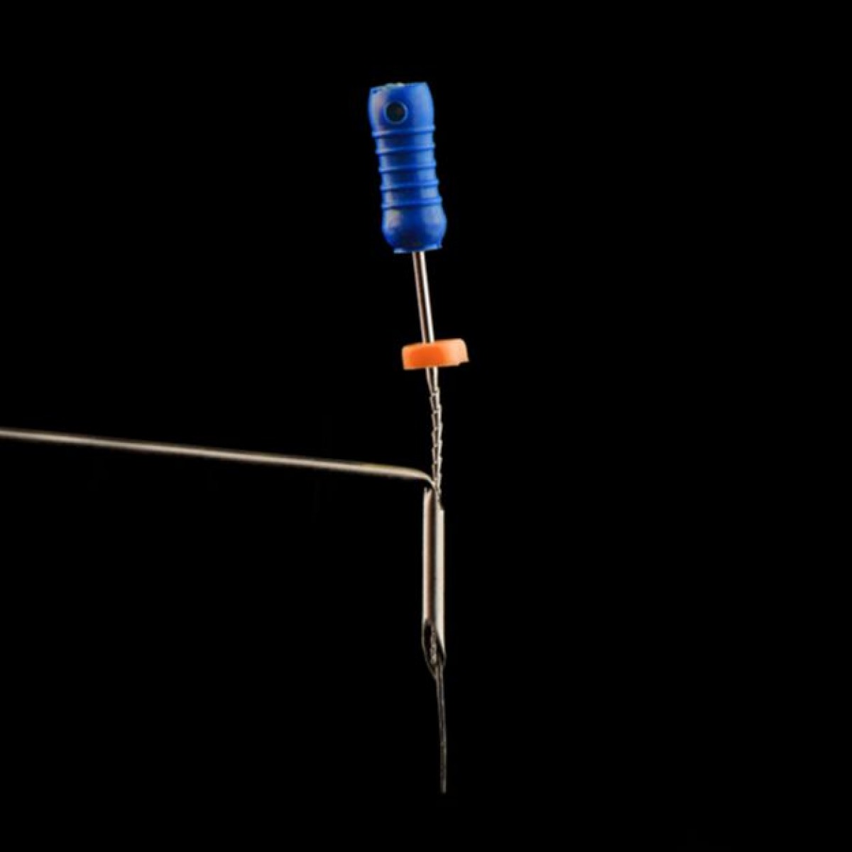
This case series reports six cases of successful removal of broken instruments using different methods including the ultrasonic, tube-and-glue, tube-and-wire, tube-and-internal shaft, and the forceps techniques and also provides a brief review of the relevant literature.
Introduction
Instrument fracture is a common occurrence during root canal treatment. The broken fragment can obstruct the root canal system; complicate irrigation, disinfection, and cleaning and shaping of the root canal system; and compromise the outcome, especially in infected teeth. Thus, prevention of instrument fracture is crucial and should take precedence over retrieval.
Hand and rotary instruments made of stainless steel and nickel-titanium (NiTi) are widely employed for cleaning and shaping of the root canal system due to their flexibility and resistance to torsional fracture. They enable achieving predictable results with nonsurgical endodontic treatment. However, instrument fracture can still occur despite adherence to the recommended protocols.
There are two approaches for the nonsurgical management of a broken instrument in the root canal system: fragment removal and fragment bypass. Optimally, the broken instrument should be removed from the root canal to facilitate effective cleaning and shaping. If nonsurgical techniques fail to remove broken instruments, surgical approaches including apicoectomy, root amputation, or intentional replantation can be used. Intentional replantation (IR) refers to the deliberate extraction and subsequent reinsertion of an endodontically treated tooth into its socket with the aim of correcting an evident clinical or radiographic endodontic failure. This method is recommended in cases where an instrument is lodged in the canal and cannot be removed with nonsurgical techniques, when a posttype crown restoration necessitates retreatment, or when performing apical surgery would result in excessive bone loss leading and a risk of damage to vital structures. Removing a broken instrument from the root canal system is complicated and requires specialized training and experience, as well as a deep understanding of the available methods, techniques, and equipment. The success of the removal procedure depends on several factors including the location, visibility, size, length, and type of the fractured instrument, as well as the curvature and radius of the root canal. Experience and fatigue of the operator are also important factors that can affect the outcome.
Many devices, techniques, and methods are available for the broken instrument retrieval. In almost all cases, the retrieval procedure consists of two main phases: initially, the root canal is prepared using rotary or ultrasonic instruments to dislodge and loosen the broken fragment and expose the coronal portion for the subsequent step. Next, specialized devices or ultrasonic techniques are employed to remove the separated instrument.
Application of ultrasonic tips is an effective method for broken instrument removal from the root canal and is utilized in almost all retrieval procedures with the success rate varying from 33% to 100%. However, even if the broken fragment in the canal becomes loose and mobile, the removal phase could be challenging. On the other hand, in some cases, the broken fragment remains immobile, and excessive ultrasonic use can result in excessive dentin removal, increasing the risk of iatrogenic errors such as transportation and perforation. Therefore, other methods should be considered for instrument removal.
Various methods have been devised for broken instrument retrieval. Application of these methods together with the use of a dental microscope may enable successful removal of the broken instrument with minimal dentin removal.
This case series reports six cases of successful removal of broken instruments using different methods and also provides a brief review of the relevant literature.
Authors: Mohsen Aminsobhani, Nasim Hashemi, Fatemeh Hamidzadeh, Pegah Sarraf
Source: https://onlinelibrary.wiley.com/
 Related articles
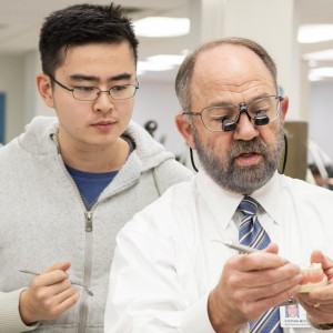
Endodontics 06 May 2022
How to manage broken endodontic instruments - Interview with Dott. Castellucci

Author: Arianna Bianchi
The fracture of endodontic instruments during root canal treatment is an event that often worries clinicians, who risk finding themselves faced with an obstacle that is difficult to overcome, with...
Products 15 July 2024
Marion Dental Enhances Patient Care with SMART Certification for Safe Amalgam Removal

Dr. Laura Fauchier, a distinguished practitioner at Marion Dental, is proud to announce her upcoming certification in the Safe Mercury Amalgam Removal Technique (SMART).
Endodontics 13 March 2024
Removal of fiber endocanal posts: guided technique vs conventional freehand technique

When due to periapical pathologies there is a need to provide root canal retreatment in teeth treated endodontically and reconstructed with a post, the first step is the removal of the post in order...

Researchers examined the plaque removal effectiveness of a personalized 3D-printed dental plaque removal mouthguard device in a clinical trial.

The new guidelines suggest conservative methods to treat tooth decay could lead to better outcomes when used with common restorative materials like fillings or caps.
Read more
Products 23 June 2026
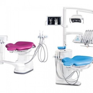
Planmeca introduces two dental units designed to support modern clinical workflows through different concepts and use cases.
Dental practice transaction can come with a variety of obstacles in the most normal of circumstances.

The American Society for Dental Aesthetics invites you to attend the 2026 Annual International Conference on Dental Aesthetics Excellence at the Conrad Nashville, October 21-24, 20
Editorials 23 June 2026
Periodontology 23 June 2026
This peer-reviewed periodontology article summarizes clinical evidence from Oral health & preventive dentistry (2026). It focuses on findings that may help dental professionals evaluate treatment...


Copyright © 2026 - All Rights Reserved
ISSN 2767-1178