

Accuracy of interdental cavity measurements performed on digital radiographs
from Dental Cadmos

Objectives: The aim of the present study was to evaluate the accuracy of linear radiographic measurements (taken on radiographs obtained with three different systems - analogic; photo-stimulable phosphor plate, PSP; and charge-coupled devices, CCD) of artificial interdental cavities.
Materials and methods: For the study, 9 posterior teeth extracted for periodontal reasons with intact crown and roots were used. Each tooth was mounted in a plaster support. At the mesial and distal aspect of each crown, two artificial cavities (one on each side) were made using cylindrical burs of different diameters, which were mounted on a calibrated milling machine. The study was conducted on a total of 18 artificial interdental cavities. For each tooth, radiographs were then taken with each of the following radiographic systems: analogic, PSP, and CCD. Two radiographs were taken with each system: one with the interposition of plexiglass between the X-ray tube and the radiographic device to simulate the presence of perioral soft tissues, the second without the interposition of plexiglass. Analogic radiographs were digitized. Each digitized analogic image as well as each radiographic image obtained with either PSP or CCD systems was calibrated. On each image, a calibrated and blind operator measured the apico-coronal (height) and mesio-distal (depth) dimensions of each cavity.
Results: The study showed that: (i) linear measurements of artificial interdental cavities of known size performed on radiographs taken with the three investigated systems are characterized by low average errors (within 0.3 mm); (ii) the magnitude of the variability of these errors may be clinically relevant, exceeding 0.5 mm and, therefore, representing a relevant aspect in identifying the most accurate system; (iii) CCD system is characterized by a non-significant tendency to show higher accuracy than the other two systems.
Conclusions: Similar accuracy was observed for linear measurements of artificial interdental cavities performed on radiographs taken with the three investigated systems (analogic, PSP, CCD).
Authors include: Francesco Malaguti, Veronica Chinellato, Jessica Miola, Alberto Borella, Roberto Farina
 Related articles
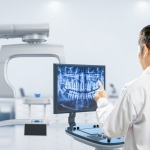
Digital radiographic imaging systems have undergone tremendous improvements since their introduction.
Orthodontics 08 October 2025
The field of orthodontics in its new era is venturing ahead to more up-to-date technological point of view.

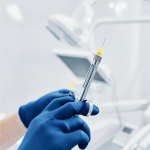
A new dual-cured resin sealer has recently been proposed as an innovative endodontic filling material.
Editorials 24 July 2025
Pitt Dental Medicine Awarded Innovation Grant in Education Award for AI-Powered Radiograph Education
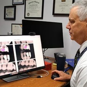
The University of Pittsburgh School of Dental Medicine Department of Restorative Dentistry and Comprehensive Care has been named the recipient of a 2025–2026 Innovation in Education Award
Restorative dentistry 30 May 2025
This clinical study was undertaken to evaluate the use of tissue-engineered bone, mesenchymal stem cells, platelet-rich plasma, and beta-tricalcium phosphate
Read more
Products 26 June 2026
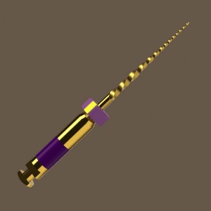
Coronal flaring is considered a key step in efficient root canal preparation, especially for difficult-to-access canals.
News 26 June 2026
The Association for Dental Safety (ADS) proudly announced the recipients of the 2026 Leadership Awards during its Annual Conference on May 27 in Salt Lake City, Utah.
News 26 June 2026

Recognition highlights the company’s doctor-led culture and continued investment in team member growth and engagement
Editorials 26 June 2026
From California to Canada: CU Anschutz School of Dental Medicine at ADEA, GRC and IADR 2026
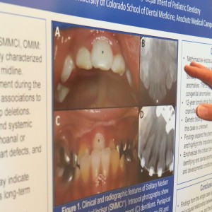
The CU Anschutz School of Dental Medicine will showcase a wide breadth of educational innovation, faculty development, clinical training and research at three major gatherings this
Oral surgery 26 June 2026
This peer-reviewed oral surgery article summarizes clinical evidence from International journal of oral and maxillofacial surgery (2026). It focuses on findings that may help dental professionals...


Copyright © 2026 - All Rights Reserved
ISSN 2767-1178