Proximal caries detection and intraoral scanner
Simona Chirico
The use of three-dimensional (3D) intraoral scanners for oral health monitoring, particularly for the detection and subsequent observation of carious lesions, is receiving increasing attention from researchers and the industry. The NIR infrared light transillumination method has been extensively studied in the literature, mainly for the detection of proximal and occlusal carious lesions. When NIR transillumination is used for proximal caries detection, NIR light is emitted directly on the buccal and lingual gingiva of the examined teeth; the light transmitted through the gum, alveolar bone and dental tissues is received by a camera that observes the teeth from the occlusal surface. With this method, the images captured show the intact enamel of a light gray color, since the NIR light is easily transmitted through this tissue, while the dentin and demineralized tissues, which diffuse more light, appear dark gray. Different NIR wavelengths were investigated in search of the best image quality for clinical application and wavelengths in the 830-1310 nm range showed better image contrast to distinguish carious lesions from tissues hard healthy teeth. The commercial device currently using NIR transillumination (DIAGNOcam, KaVo, Germany / CariVu, DEXIS, USA), as well as the prototypes used for research purposes, employ 780nm laser light, showing promising diagnostic performance for the detection of interproximal lesions. . In the prototype used in this study, efforts were made to reduce the noise (graphic distortions) observed on NIR images compared to the existing commercial device: 1) eliminating spots by using light-emitting diode (LED) laser light instead of coherent laser light e 2) employing a new design aimed at improving the adaptation of the NIR tip to the oral tissues and thus reducing the overexposed areas due to the reflected light. Other improvements of the proposed prototype, which could ensure better image quality, are the use of a different wavelength (850 nm) and the wider field of view. Finally, by combining the NIR scan with the intraoral scan, the exact position of each NIR image taken is recorded on the 3D model of the teeth.
Materials and methods
In a clinical study, soon to be published in the Journal of Dentistry, the authors evaluated the validity of an intraoral scanner system with infrared transillumination (NIR) to aid in the detection of proximal caries lesions by comparing their diagnostic performance with that of conventional methods. caries detection and with those of an intraoral camera with NIR transillumination (DIAGNOcam). Ninety-five permanent posterior teeth were examined and divided into three groups:
- Group 1 where interproximal caries were investigated using a tip prototype working with the TRIOS 4 intraoral scanner system (3Shape TRIOS A / S, Denmark) with NIR light emission
- Group 2 where interproximal caries were detected by DIAGNOcam, e
- Group 3 or control where interproximal caries have been investigated by visual and radiographic examination according to ICDAS criteria.
One or two proximal surfaces per tooth, healthy or with carious lesions at different stages, were examined (N1 = 158). Histological evaluation was used as the reference standard.
Results
All methods showed excellent intra-examiner reliability. Two independent examiners evaluated the NIR images obtained with both devices. The agreement between the two examiners, who evaluated the NIR images, was substantial. The intraoral scanner and the DIAGNOcam showed similar diagnostic performance. Regarding the initial carious lesions, the evaluation of the NIR image resulted in equal or better sensitivity than the radiographic evaluation and superior to the visual examination. The radiographic image and the NIR evaluation led to similar performance in detecting moderate-extensive dentinal carious lesions, while the visual examination showed a lower value.
Conclusions
From the data of this study, which must be confirmed in other similar studies, it can be concluded that the intraoral scanner system with NIR transillumination and the DIAGNOcam guarantee a good overall diagnostic performance for the detection of interproximal caries. Conventional caries detection methods, on the other hand, have shown lower sensitivity in the early stages of carious lesions.
For additional information: Intraoral scanner featuring transillumination for proximal caries detection. An in vitro validation study on permanent posterior teeth.
 Related articles
Products 26 August 2025
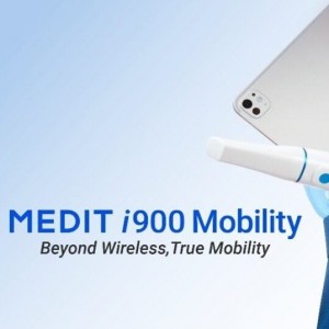
Medit, a global leader in digital dentistry, today introduces the Medit i900 Mobility, a next-generation intraoral scanner designed to deliver simplicity and true mobility to clinical workflows.
News 01 April 2025
OMNIVISION, a leading global developer of semiconductor technology—including advanced digital imaging, analog, and display solutions—and Biotech Dental, a specialist in the design and manufacture...
News 06 February 2025
Technavio Forecasts USD 915.75 Million Growth in Intraoral Scanners Market by 2028
The global intraoral scanners (IOS) market is poised for significant growth, with an estimated increase of USD 915.75 million from 2024 to 2028, reflecting a CAGR of 11.68%, according to Technavio.
Products 06 January 2025
Technavio Report: AI and Tech Advances Propel Intraoral Scanners Market Expansion
The global intraoral scanners market is projected to grow by USD 915.75 million between 2024 and 2028, expanding at a compound annual growth rate (CAGR) of 11.68%, according to Technavio in a recent...
Medit, a global leader in intraoral scanning technology, is making a significant impact in the Slovenian market as it continues its expansion across the Balkans.
 Read more
Oral pathology 25 November 2025
Virtual microscopy (VM) is a technology for showing microscope slides using computers and could be considered a progression of classic methodology using optical microscopes.
For every assist this season, the insurance provider will donate $25 to TUSDM Cares for Veterans
Products 25 November 2025
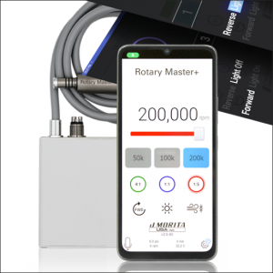
J. MORITA USA, a world leader in handpiece technology, has announced the Rotary Master+ Electric Motor. Compatible with Morita TorqTech electric attachments and most competing electric handpieces on...
News 25 November 2025
Let’s be honest: nothing kills the vibe quite like bad breath. However, while 85% of people prefer for someone to tell them if their breath needs some freshening up, only 15% are willing to break...
News 25 November 2025
Vitana Pediatric & Orthodontic Partners (Vitana), a dentist-led dental partnership organization (DPO) focused exclusively on elite pediatric dental and orthodontic practices with operations in...


