

Restoration of endodontically treated molar: a clinical case
Authors: Dr. Martignoni, Prof. Grandini

The restoration of the endodontically treated molar has always been a debated topic. The clinician faces many different challenges and it is important to know how to approach every single patient depending on the different clinical situations.
Recently, an evidence-based treatment planning for the restoration of endodontically treated molar has been proposed (reference: Evidence-based treatment planning for the restoration of endodontically treated single teeth: importance of coronal seal, post vs no post, and indirect vs direct restoration).
The importance of the coronal seal is undisputable for the long term success of the endodontic treatment.
The clinician has to decide if a particular tooth needs a post or not, if a direct or an indirect restoration is required, which material needs to be used. In cases where there is enough tooth structure left, and the patient has no parafunctional habits, direct restoration can be used to reduce the amount of tooth structure that needs to be removed for the preparation of the indirect restoration.
The following case is an example of a molar, with four sound remaining walls, that is directly restored after the root canal treatment.
The patient, 26 years old, was referred for an endodontic treatment after a pulpitis occurred on the tooth 2.6 and an emergency treatment was carried out by another dentist (rx 1)

Rx 1: Periapical rx of 2.6
After local anesthesia, the rubber dam is placed to isolate the operating field. The temporary restoration is clearly visible (fig 1).
After removing it, the canal orifices are located (fig 2), including MB2 and the shaping and irrigation is performed (fig 3).
A sequence of ProTaper Gold instruments is used to shape the root canals and the endodontic space is obturated and sealed with the continuous wave of condensation technique.

Fig 1: temporary restoration

Fig 2: canal orifices are located

Fig 3: shaping is performed
Fig. 4 shows the result after the floor of the cavity preparation has been cleaned using AH Cleaner, to remove all remnants of the cement and to be prepared for the next steps of the adhesion.

Fig.4: finishing of endodontic treatment
Selective etching of the enamel (fig 5) is one of the possibilities we have when using a universal adhesive system. Enamel is a substrate that benefits from the etching phase, while on dentin results are quite similar in both etch and rinse and etch and dry mode.

Fig 5: selective etching of enamel
After the Dentin Bonding Agent has been light cured, a bulk filling material (fig 6) is used to replace the missing dentin structure. Bulk fillers can be used in 4mm increments and give us a few clinical advantages, being easy to use and requiring a very limited time.

Fig 6: bulk filler
The last layer when using this “bulk and body” technique is the so called capping layer. 2mm of resin composite are used to rebuild the occlusal anatomy. Fig 7 and 8 show the modelling of the cusps, until the restoration is completed (fig 9).

Fig 7: modelling of cusps

Fig 8: modelling of cusps

Fig 9: restoration is completed
A careful finishing and polishing is carried out, leading to a correct integration of the restoration in the remaining tooth substance (fig 10).

Fig. 10: restoration is completed
 Related articles
Prosthodontics 23 October 2019
Rehabilitation of endodontically treated molars: is better to choose endocrown or crown with post?

The restoration of endodontically treated teeth is always a topic of crucial attention for dentists. If until a few years ago, the therapeutic choice of...
Read more
USA 19 July 2026 - 30 November -0001
ADA Scientific Session 2026 in Indianapolis: Event Preview and Professional Highlights

The ADA Scientific Session 2026 will take place from October 8, 2026 to October 10, 2026 in Indianapolis, IN, USA, offering dental professionals a focused environment for continuing education,...
Edra Professional Books 18 July 2026
The Complete Book on Dental Marketing: A Practical Reference for Dental Professionals

The Complete Book on Dental Marketing is an Edra professional dentistry reference focused on clinical practice, education and treatment planning.
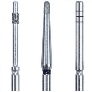
Eagle Crown Lengthening Burs are designed to make surgical precision effortless—helping clinicians expose more tooth structure smoothly, efficiently, and with total control.
News 17 July 2026
The new Bogotá, Colombia facility strengthens Roland DGA’s long-standing commitment to dental professionals and partners across the region.

New integration streamlines patient financing within CareStack’s practice management platform, making it easier for providers to help patients move forward with care.


Copyright © 2026 - All Rights Reserved
ISSN 2767-1178