


Immediate loading implants with one step technique and double milling technique
Authors: F. Zingari F. Gallo S. Ghezzi E. Carozzi
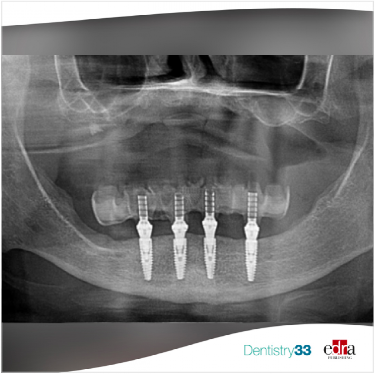
A 70 years patient, in good general condition, came to our attention as she was wearing incongruous mobile prostheses, both upper and lower.
At radiographic control with orthopantomography and CBCT, it is evident that both the implants in the upper and lower arch are affected by peri-implantitis.
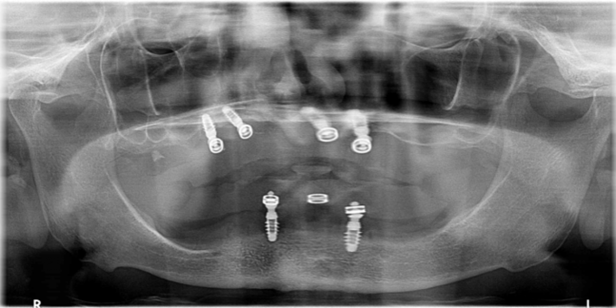
TREATMENT PLAN
It was decided to plan, first of all, an extraction of the present implants and a subsequent implant-prosthetic rehabilitation with immediate loading, initially of the lower arch and then of the upper one too.
PRE-SURGICAL PHASE
The therapeutic project begins with the relining of the patient's old prostheses, which will then undergo intraoral scanning for the creation of new prostheses that will allow us to rehabilitate the vertical dimension lost over time, before proceeding to surgery and prosthetic rehabilitation.
In the upper arch, the removal of the existing implants is carried out before the creation of the new prosthesis, while in the lower arch, since there is only one implant, it is decided to extract it at the time of implant surgery.
Once the old prosthesis of the patient have been scanned, the laboratory will be able to make prototypes that will be tested in the mouth. Once the prototypes are satisfactory, the laboratory continues with the creation of the upper and lower total dentures.
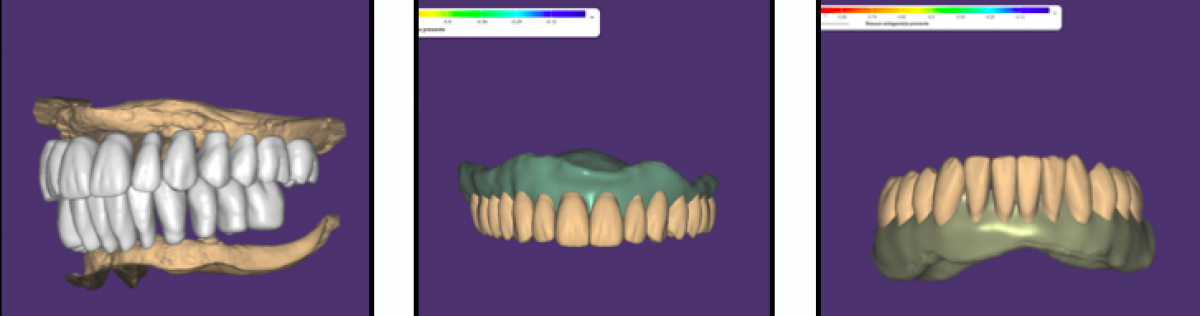
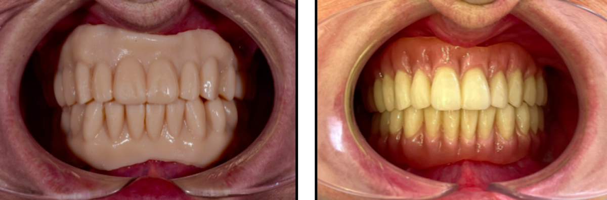
The patient will wear the new prostheses for about two months, to get used to the new vertical dimension, once the patient does not report articular disorders we can proceed with the surgical act, which will involve the placement of 4 implants in the lower arch with subsequent immediate loading.
SURGICAL PHASE
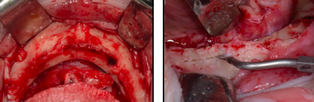
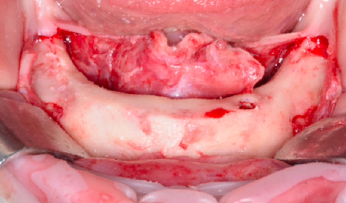
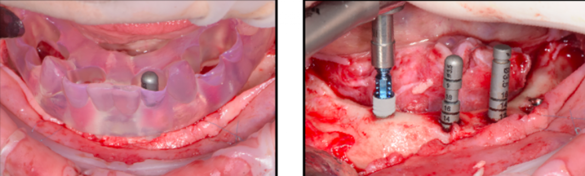
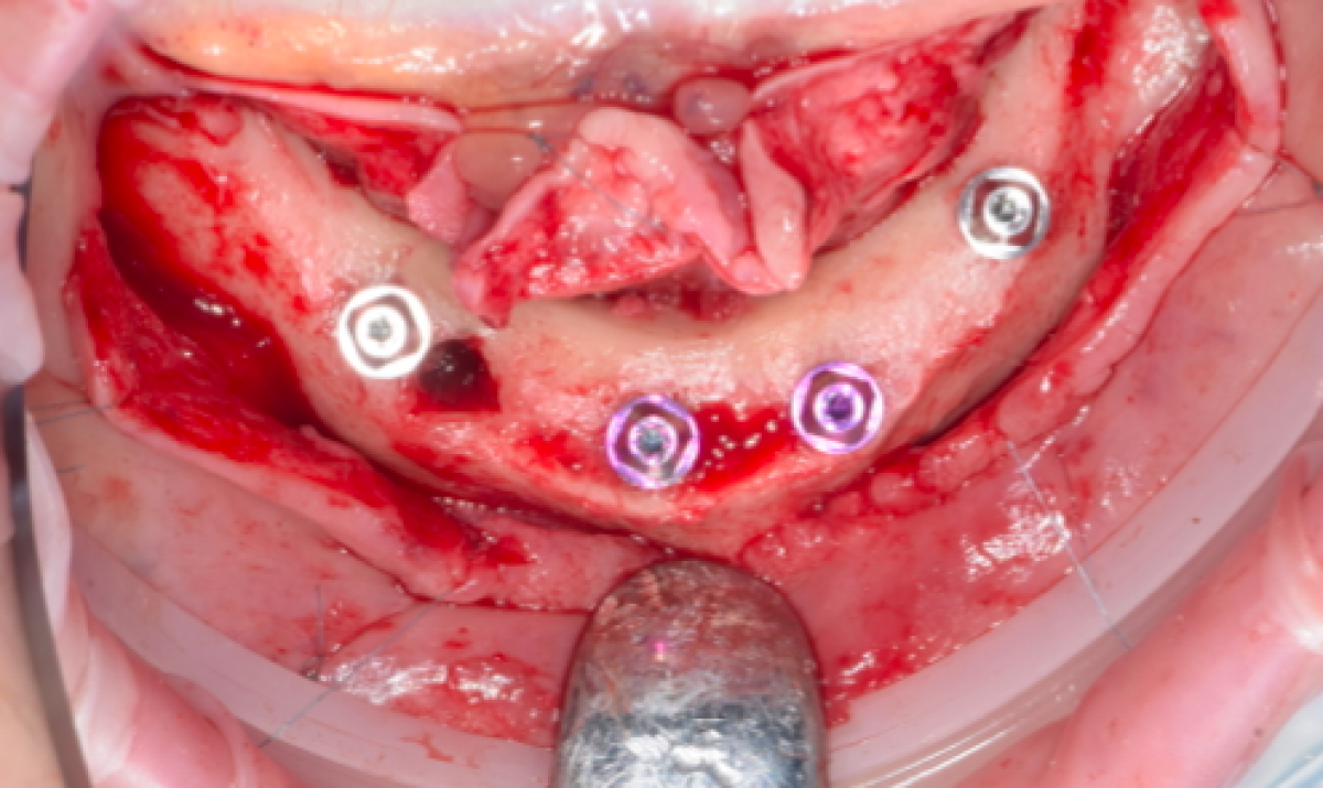
The surgical phase involves, first of all, the extraction of the implants affected by peri-implantitis, still present in the lower arch in region 33 and 43. A preoperative incision is then made from region 37 to 47 with distal incisions, and after an accurate skeletonization of the maxilla, an ostectomy of the residual alveolar ridge is performed, to obtain a mandibular regularization, using SonicFlex. With sonic instrumentation the cutting action is achieved through the oscillation of a tip that vibrates at sonic frequencies in the range of 5,000-9,000 Hz, the controlled movement, precision, selective cutting on hard tissue and ergonomics of this instrument allows for effective results with minimal invasiveness. This procedure will make prosthetic rehabilitation more appropriate both cosmetically and functionally, and will facilitate patient cleanability of the prosthesis.
PROSTHETIC PHASE
The prosthetic phase begins with the detection of the implant position by means of the One Step technique, i.e. using the template used as analogical guide during surgery. The latter is perforated in the points of exit of the implants, to make room for the turrets that once screwed to the Mua are fixed to the template with Pattern resin, this will allow the laboratory to have the exact position of the implants compared to the prosthetic rehabilitation that the patient is already wearing and therefore already functionalized.
Once the lab receives the template with turrets, it is ready to prepare the immediate load, which is done with the "Double milling" technique.
This technique consists, as a first step, in drawing the anatomical part on the CAD, using the diagnostic wax-up, which in this case is obtained by scanning the existing prosthesis. At this point it is possible to design the sub-structure that will act as a support for the prosthesis in immediate load. The sub-structure is generated by subtraction starting from the anatomical project. In this way, the future denture teeth and the connections between them and the linkers are structurally well supported. The files of the sub-structure thus designed are sent to the Cam software where the nesting phase takes place, which consists in positioning the file of the sub-structure project inside the virtual disc. Once the structure is milled as designed, it is not detached from the disc but is filled in its empty spaces with a mixture of PmmA composed of dentin enamel and transparent. The last step involves the insertion of the disc obtained in this way in the milling machine in order to carry out the second Cam phase, thus realizing the anatomical part of the prosthetic artifact designed. The "double milling" technique allows us to obtain both the sub-structure and the anatomical part perfectly identical to the project and joined together without interruption.

 Read more
Europe 27 September 2026 - 30 November -0001
Digital Dentistry Society Global Congress 2026: Event Preview and Professional Highlights

The Digital Dentistry Society Global Congress 2026 will take place from October 22, 2026 to October 24, 2026 in TBD, Rotating Europe, offering dental professionals a focused environment for...
Editorials 27 September 2026
Dentistry33 survey highlights replacement care, deep caries and evolving restorative workflows
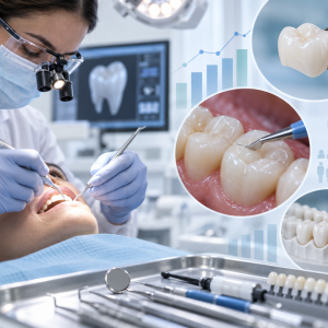
Reader survey finds direct composites remain dominant, secondary caries is the leading reason for restoration replacement, and clinicians want more training in pulp preservation
Reader survey finds direct composites remain dominant, secondary caries is the leading reason for restoration replacement, and clinicians want more training in pulp preservation
Edra Professional Books 26 September 2026
Manual of oral surgery: A Practical Reference for Dental Professionals
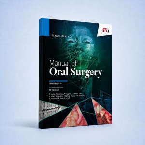
Manual of oral surgery is an Edra professional dentistry reference focused on clinical practice, education and treatment planning.
Products 25 September 2026
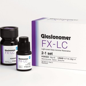
Shofu Dental’s line of dental restorative materials now includes FX-LC, a next-generation, light-cure, resin-reinforced glass ionomer formulated to deliver high strength, efficient 4
News 25 September 2026

The scholarships are part of an ongoing workforce solutions strategy to help address the need for skilled dental hygienists



Copyright © 2026 - All Rights Reserved
ISSN 2767-1178