


Three-Dimensional Facial Scanning in Dentistry: Why and How?
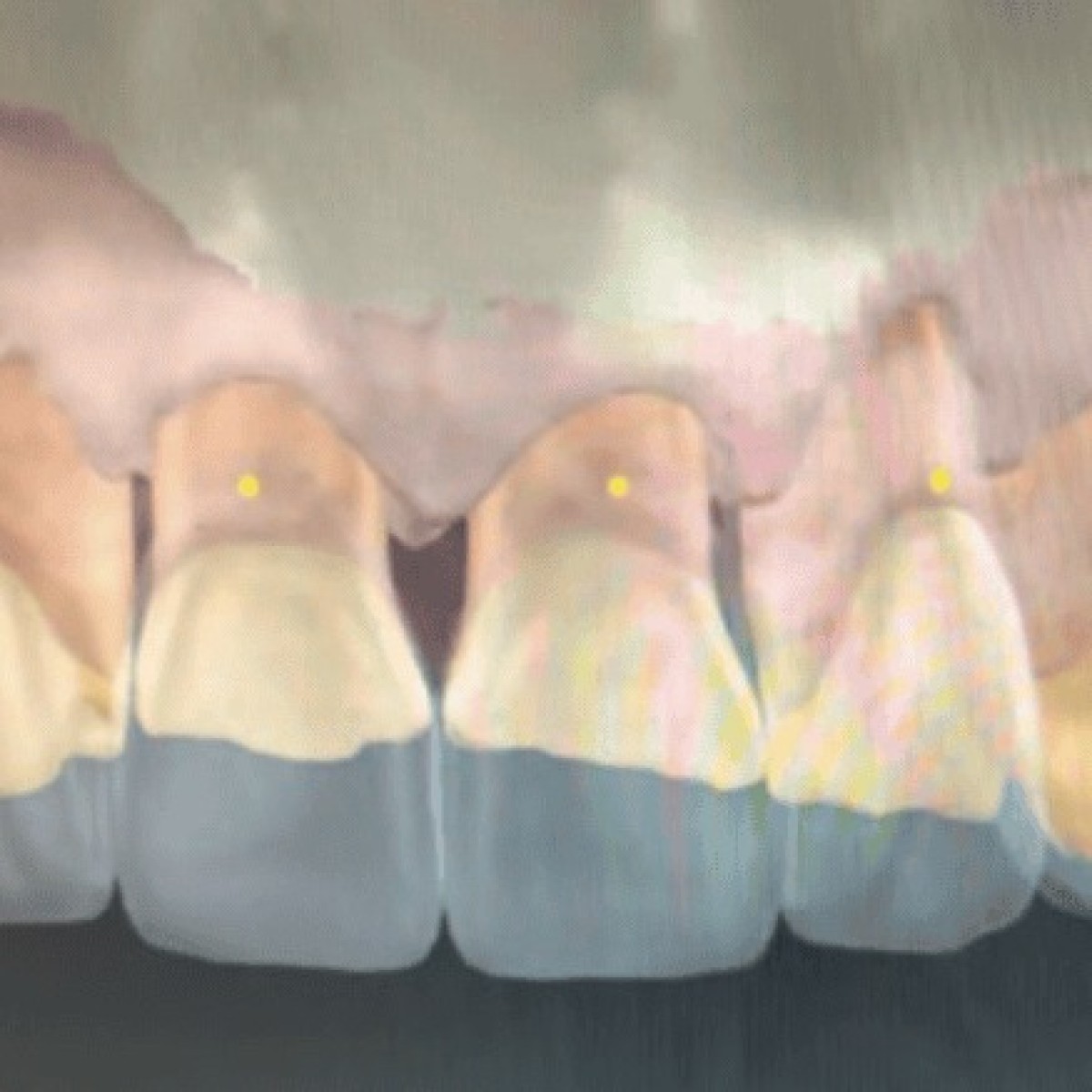
This technology allows the clinician to visualize the patient’s face in a digital format in order to establish the ideal central incisor position and incisal edge position, buccal corridor, and establish an ideal horizontal plane for facial analysis and the horizontal occlusion plane.
Three-dimensional facial scanning has been utilized in dentistry for the past 7 years. This technology allows the clinician to visualize the patient’s face in a digital format in order to establish the ideal central incisor position and incisal edge position, buccal corridor, and establish an ideal horizontal plane for facial analysis and the horizontal occlusion plane. 1-6
This is especially important for denture anterior tooth setups, All-on-X implant cases, smile designs, and provisional restorations.
The prosthodontist, restorative dentist, or dental surgeon can use this technology, in coordination with the laboratory, to create the ideal tooth position based on the patient’s lips and face. AI analysis of patient facial angles allows the design of the teeth to harmonize with the patient’s face, eyes, nose, ears, lips, cheeks, chin, and forehead in order to create the ideal tooth position. The AI processes in this software align asymmetry and facial lines to guide evaluation and accurate tooth setup. Understanding that all patients differ in these variables, obtaining an ideal facial maxillary face-bow transfer is mandatory. 7-9
Each patient’s facial characteristics are unique. This is critically important when creating smiles and determining tooth position, particularly in All-on-X cases, where the midline, maxillary cant, and plane of occlusion must all be ideal preoperatively, during surgery, and at postoperative prosthetic delivery (Figure 1).
When designing the ideal smile and tooth position for a denture patient, it is critical to use 3D facial scanning to establish the correct midline, facial lip support, and buccal corridor alignment so that the maxillary cant and the facial anatomical structures are all in alignment and appear natural in both incisal position and tooth length (Figure 2). Selecting tooth size and shape is a highly subjective step in building patient and dentist confidence. Utilizing digital tooth selection saves time, creates perfect proportions, and is easy to teach to dental professionals and dental students. In addition, the 3D facial scan must be able to acquire the facial scan and simultaneously estimate and generate a reference plane of occlusion for virtual articulation (Figure 3).
The analog method of fabricating dentures is adequate, but the digital format is much easier to perform and far better suited for student education. Tooth selection guides are much easier to use in a digital format than a physical palette of various sizes and lengths. Also, 3D facial scanning can determine tooth position within minutes, whereas searching for the correct teeth on an analog palette can be time-consuming, difficult to interpret, and highly subjective. Utilizing digital technologies, we can select the teeth within minutes and formulate the ideal proportions based on the facial AI analytics that are now available. This can then be transferred to a PMMA digital-printed model for a try-in visit. Confirming occlusion, central length, style of teeth, smile display and ideal position for lip support is now an easy step without the wax getting warm, soft, and unstable (Figure 4).
There are 3 types of All-on-X cases: edentulous patients, dentate patients, and patients with only a few teeth remaining. In all these scenarios, the vertical dimension of occlusion (VDO) is a critical component in designing the initial interim prosthesis on implants—the All-on-X PMMA prosthesis. The IOS scanning needs to be accurate, precise, and true from the scan to the printer. 10,11 Calibration of these scanners is necessary. The first step includes photographs, IOS scan with accuracy and precision, and face-bow transfer (Artex or KDFA [Kois Dento-facial analyzer]). In addition, natural head position is an important clinical reference when establishing facial orientation (Figure 5). 12
When treating aesthetic anterior implant cases and evaluating the CEJ area for an ideal provisional to capture cervical anatomy, CBCT scanning is paramount. 13 However, when treating full arches, this cervical component becomes more critical for FP1 implant cases. If the patient treatment is being planned for FP3 cases, the 3D facial scan becomes more relative together with the CBCT scan.
Utilizing facial aesthetics and 3D facial analysis, the design of the implant positions involves both surgical and laboratory components; once those positions are determined, the restoration must align with the face (point-facial-marker-merger). Midlines are critical and, in many cases, are not ideal when placed on the patient. A maxillary cant can also be unaesthetic or malpositioned, which is very frustrating for the restorative dentist attempting to establish occlusion with the mandibular arch. 7,12 When positions are incorrect, the restorative procedure can be extended by 4 to 5 hours. Predetermining ideal positions, midlines, buccal corridors, incisal edge, central lengths, and dimensions using the 3D facial scanner minimizes errors and supports ideal delivery of prosthetic restorations on implants.
During All-on-X procedures, surgeons have specific sites and bone areas for implant placement. Making the final digital impression utilizing photogrammetry technology with IOS scanner (Aoralscan Elite [SHINING 3D) can now be performed in 3 to 4 minutes.
This has improved the efficiency and accuracy of the impression appointment, creating an accurate master model (Figure 6).
When combined with laboratory work and prosthetic restorations, these 3 specialists must coordinate to deliver the proper prosthesis in a timely manner; keeping a patient under surgical access for more than 2 to 3 hours is not recommended. Using 3D facial scanning, we can deliver a predictable position of the anterior and posterior occlusion, along with condylar analysis, for every patient. Condylar position must be confirmed with CBCT scan (Vatech) from the first appointment, to the PMMA provisional, to the final try-in prostheses, to the final restoration. This is a critical factor in understanding and confirming that the VDO is in the correct zero position (Figure 7).
It has been suggested that face-bow transfers may not be necessary to obtain the correct alignment of the maxilla-mandibular position. With over 30 years of experience, we have observed that, although 3D facial scanning can bring us very close, a 3D facial face-bow transfer is still necessary to achieve the ideal position. Skipping this step may result in unbalanced midlines, occlusions, and maxillary cants. Understanding the horizontal plane, merging of the photographs and 3D facial scans all play a role in minimizing distortion of the merging transfer. The final restoration will be a titanium frame directly to the MUA abutments and a zirconia ceramic screw-retained final implant restoration (Figure 8).
Since 2012, we have performed every smile design case we have treated with a 3D facial scan, a CBCT scan, and digital design software. In the past 5 years, 3D facial scanning has become a priority. With this technology, we can show the patient an ideal simulation, along with virtual AI videos and simulations of their smile (PreVu [PreVu Dental]) (Figure 9). 4
Because the teeth are already in position and we are simply adding ceramic or provisional material, 3D facial scanning technology, in addition to intraoral (IOS) scanning, photographs, facebow transfer, videos, and CBCT scanning, has allowed us to perform this treatment in a very predictable manner with minimal adjustments to aesthetics, occlusion, and maxillary cant (Figure 10).
We used a 3D facial scanner (MetiSmile [SHINING 3D]) that allows a full 3D facial scan simulation and facial analysis to be completed in 2 to 4 minutes. It is recommended that the patient wear no facial garments and the ears remain uncovered so the full AI component can be utilized. Glasses and headwear must also be removed before the 3D facial scan.
The digital workflow proceeds as follows:
Once the clinicians receive the data file, they can confirm the completed tooth positions: the midline position, lip support, facial support, buccal corridor, occlusal design, VDO, and the ideal position of the temporo-mandibular joint (TMJ) during any and all mandibular movements. This is the treating dentist’s sole responsibility…this serves as the most important quality control.
This software also offers an option to incorporate mandibular movements (MetiSmile-MR Jaw tracking [SHINING 3D]) through the facial scan. This may take 20 to 30 minutes to produce, but it can be extremely valuable for patients with severely worn dentition or those who have lost their VDO. Utilizing “Shimabashi” measurements (CEJ-to-CEJ) for occlusion, we can restore the VDO of this patient predictably with the FMR concepts; a-Tripod anterior aesthetically driven b-bio-esthetic anatomical positioning to restore VDO c-TMJ verification of VDO. In addition, posterior contacts confirm new VDO inMIP position (Figure 13).
Once the dentist approves the files, they are sent to the printing laboratory—either an in-office printer or a laboratory printer—to produce one of the following:
After final preparation and provisionals are completed, the patient is scanned for final restorations (Figure 14). An additional scan of the provisional restorations is made to copy the ideal occlusion, canine guidance, and incisal edge position, as well as all aesthetic aspects of the newly formed smile. This is critical to capture and copy the contours, lengths, shapes, and occlusal morphology of the provisionals to the final restorations. The final materials chosen for this patient were feldspathic ceramics for the anterior teeth, an IPS e.max (Ivoclar) posterior fixed bridge on the maxillary right side, and individual IPS e.max crowns on the maxillary left side (Figures 15 to 17).
Many digital files are available today, and the AI components of these programs have significantly reduced the time required for design and execution (MetiSmile). Obstacles for most clinicians may include financial constraints, access to training, or the comfort of familiar routines and the redundancy of doing things simply because they feel comfortable. After 35 years in dental education, I have learned one essential lesson in the dental profession: learning new technologies is a must. Stepping out of one’s comfort zone is valuable; doing things merely because they have always worked wastes a great deal of time and does not elevate our profession. Our profession is constantly changing, and as our patients’ demands increase, our students’ thirst for knowledge, and our ability to be better clinicians, will always be the driving force of the digital dental world; as well as the dynamics of digital restorations. I encourage all our younger colleagues to embrace technology and commit to learning new systems and new software to help grow the future of dentistry.
In conclusion, 3D facial scanning is highly predictable, and any staff member or student can be trained to use it within half a day. It is a valuable tool for the laboratory, the surgeon, and the restorative dentist. The software currently available allows data files to be easily transferred and merged across multiple laboratories. The data acquisition and output are dramatically faster and more efficient than ever before. Facial scanning integrated with AI delivers exact tooth positioning and occlusal planning, reducing multiple try-ins and guesswork in denture and prosthetic seating. Utilizing 3D facial scanning will reduce hours of clinical chair time and streamline the digital workflow of any institution, private office, or group practice (Figure 18). 14,15
The author wishes to thank Carey Lyons, Lana Colantuono, Tory Perry, Norelis Tigres, Lou Defilipppo, and Steve Kim MDT (Wveneer).
1. Salazar-Gamarra et al; accuracy of mobile devices compatible with 3-D scanners for facial digitization systematic review; JMed Internet Res; 2020:22(10)
2. Pellitteri et al. Comparison of the accuracy of digital face scans obtained by two different scanners Angle Orthod. 2021;91 (5):641-49.
3. D’Ettorre, et.al. A comparison and smart phones structured like technology three dimensional facial scanning; Angle Orthod 2022;92(3);358-63
5. Accuracy of 3D facial scans: a comparison of three different scanning system in an in vivo study
6. Federica Pellitteri 1, Fabrizio Scisciola 2, Francesca Cremonini 2, Matilde Baciliero 2, Luca Lombardo; Prog Orthod. 2023 Dec 25;24(1):44.
7. Factors that influence the accuracy of maxillomandibular relationship at maximum intercuspation acquired by using intraoral scanners: a systematic review Revilla-León M, Barmak AB, Tohme H, Yilmaz B, Kois JC, Gómez-Polo M. J Dent. 2023;138:104718.
8. An in vitro 3D evaluation of the accuracy of 4 intra-oral scanners on a 6-implant model. Sami T, Goldstein G, Vafiadis DC; Journ Prosth Dent.2020 Dec;124(6):748-754
9. An overview of the digital occlusion technologies: intraoral scanners, jaw tracking systems, and computerized occlusal analysis devices M.Revilla-León M, Kois DE, Zeitler JM, Att W, Kois JC.. J Esthet Restor Dent. 2023;35(5):735-744.
10. An in vitro 3D evaluation of the accuracy of 4 intra-oral scanners on a 6-implant model. Sami T, Goldstein G, Vafiadis DC; Journ Prosth Dent.2020 Dec;124(6):748-754
11. Evaluation of trueness and precision of 3 face-scanning devices. Bor S., Oğuz F., Özdemir D. Am. J. Orthod. Dentofac. Orthop. 2025;168:358–366.
12. An overview of the different digital facebow methods for transferring the maxillary cast into the virtual articulator Marta Revilla-Leon, Jonathan M. Zeitler, John C Kois; JERD 2024 Dec; 36(12):1675-1686
13. Immediate Implant placement of a Single Central Incisor Using a Cad/Cam Crown-Root form Technique: From Provisional to Final Restoration. Vafiadis DC, Goldstein G, Garber D, Lambrakos A, Kowalski BJ. Journal of Esthetics and Restorative Dentistry. Vol 29, Issue 1, 13-21 Jan-Feb 2017
14. Predictable Full Digital Workflow Using Stackable Surgical Templates for Complete Dental Arch Rehabilitation with Implant-Supported Fixed Restorations-Case Series and Proof of Concept Cristache C.M., Burlacu Vatamanu O.E., Butnarasu C.C., Mihut T., Sgiea E.D.. Dent. J. 2024;12:347
15. Techniques and accuracy for aligning facial and intraoral digital scans to integrate a 3-dimensional virtual patient: A systematic review. Lawand G., Tohme H., Azevedo L., Martin W., Gonzaga L., Nassif M., Revilla-León M. J. Prosthet. Dent. 2025
Dr. Vafiadis received his dental degree and prosthodontic specialty training at the New York University (NYU) College of Dentistry. He is currently an associate professor of prosthodontics and the director of the Full-Mouth Rehabilitation CE course at the NYU College of Dentistry. He is an adjunct professor of prosthodontics at Nova Southeastern University College of Dental Medicine and is a Fellow of the American Academy of Esthetic Dentistry. He lectures nationally and internationally for the NYU Continuing Education department. Dr. Vafiadis has been published and has lectured on various topics, such as aesthetics, implant designs, computer restorations, ceramic materials, and occlusion. He is the founder of the New York Smile Institute in New York City. He can be reached at drdean17@gmail.com.
Disclosure: Dr. Vafiadis is a key opinion leader for SHINING 3D, Vatech, Ritter, and FMR Education.
Dr. Dean C. Vafiadis will host a FREE CE WEBINAR on December 9 at 8 PM (EST) that follows up on the content of this article.
Source: https://us02web.zoom.us/webinar/register/WN_Qnq786gKT_C2R1Feh0C2eA#/registration
 Related articles
Orthodontics 02 April 2025
CBCT in orthodontics: assessment of treatment outcomes and indications for its use

Since its introduction into dentistry in 1998, CBCT has become increasingly utilized for orthodontic diagnosis, treatment planning and research.

Authors: Prof Dr Daniele Cardaropoli, Dr Alessandro Roffredo
First of 3 clinical cases in which three-dimensional radiology has allowed us to avoid making diagnostic errors otherwise not evaluable with two-dimensional radiography alone.
Oral surgery 15 October 2026
This peer-reviewed oral surgery article summarizes clinical evidence from International journal of oral and maxillofacial surgery (2026). It focuses on findings that may help dental professionals...
Oral surgery 07 October 2026
This peer-reviewed oral surgery article summarizes clinical evidence from International journal of oral and maxillofacial surgery (2026). It focuses on findings that may help dental professionals...
Oral surgery 25 September 2026
This peer-reviewed oral surgery article summarizes clinical evidence from International journal of oral and maxillofacial surgery (2026). It focuses on findings that may help dental professionals...
Read more
News 02 November 2026

Partnership adds three family dental practices in West Bend, Jackson, and Saukville, extending First Choice Dental’s presence in southeastern Wisconsin
News 02 November 2026

Registration extended through Monday, November 2, 2026, for the November 12–14 retreat at The Ballantyne in Charlotte, North Carolina.
Editorials 02 November 2026
Texas A&M College of Dentistry’s talent shows have long served as opportunities for students to show off some of their artistic talents.
Oral Hygiene & Prevention 02 November 2026
This peer-reviewed oral hygiene & prevention article summarizes clinical evidence from Odontology (2026). It focuses on findings that may help dental professionals evaluate treatment decisions,...
Europe 01 November 2026 - 30 November -0001
Osteology Symposium 2027: Event Preview and Professional Highlights

The Osteology Symposium 2027 will take place from April 22, 2027 to April 24, 2027 in TBD, Rotating Europe, offering dental professionals a focused environment for continuing education, clinical...



Copyright © 2026 - All Rights Reserved
ISSN 2767-1178