

Diagnosis and treatment planning of orthodontic patients with 3-dimensional dentofacial records
Introduction
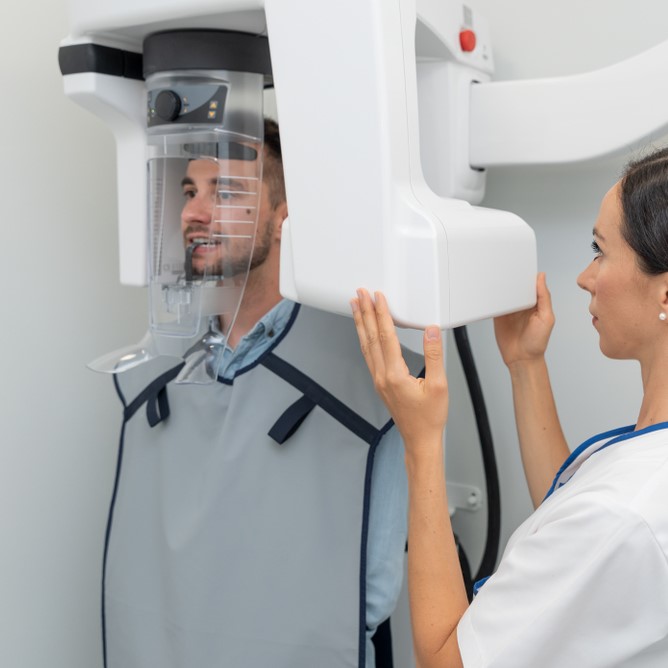
Cephalometrics has been the foundation of orthodontic diagnosis for many years. However, for many orthodontic patients, a lateral cephalogram might not be necessary. The aim of this study was to compare the diagnosis and treatment planning agreement between standard records and nonradiographic 3-dimensional (3D) dentofacial photogrammetry records.
Methods
Twenty patients had standard orthodontic records taken for their treatment as well as extraoral and intraoral 3D images. Twelve evaluators examined the standard records and then completed diagnosis and treatment planning questionnaires. They repeated the process 4 to 6 weeks later by using 3D photographic images along with the panoramic radiographs. Each evaluator also evaluated 2 random orthodontic cases twice with each method to evaluate consistency within each method. At the end of study, each evaluator was asked to complete a survey to document his or her experiences with the 3D photogrammetry method. Descriptive and kappa statistics were used to determine the agreement.
Results
Most diagnosis parameters had fair agreement between the methods and within each method. Skeletal and dental relationships had excellent agreement between and within the methods as well as most treatment decisions such as the need for extractions and surgery. Most evaluators (91.7%) thought that cephalometric x-rays would be needed only some of the time in diagnosis and treatment planning. Most evaluators (83.33%) thought that cephalometric radiographs are not needed in patients with a Class I ± a quarter cusp with crowding or spacing.
Conclusions
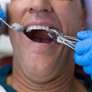
Most diagnostic decisions had fair agreement within and between the 2 methods. The decision to extract and the need for orthognathic surgery had excellent agreement between the cephalometric and photogrammetric methods. The majority of examiners agreed that patients with Class I malocclusions ± a quarter cusp with no obvious skeletal discrepancy can be diagnosed and planned without a cephalometric radiograph.
Authors: Amornrut Manosudprasit, Arshan Haghi, Veerasathpurush Allareddy, Mohamed I. Masoud
Source: https://www.sciencedirect.com/
 Related articles
This study was not funded by any organization or institution or any research grant company.
Digital Dentistry 30 September 2025
Diagnosis and Treatment Planning for Predictable Restorative Outcomes
The dilemma in comprehensive dentistry is that dentists are often focused on restoring teeth for esthetic outcomes, and if occlusion is not taken into account during diagnosis and treatment planning,...
Periodontology 25 September 2025
Throughout history, education has evolved, and new teaching/learning methods have been developed.
Periodontology 10 September 2025

To update the findings of a systematic review from the year 2016 on the evidence for the accuracy and potential benefits of cone beam computed tomography (CBCT) in periodontal diagnostics.
Orthodontics 08 September 2025
Records needed for orthodontic diagnosis and treatment planning: a systematic review
Traditionally, dental models, facial and intra-oral photographs and a set of two-dimensional radiographs are used for orthodontic diagnosis and treatment planning.
Read more
USA 12 July 2026 - 30 November -0001
DSO Leadership Summit 2026 in Austin: Event Preview and Professional Highlights

The DSO Leadership Summit 2026 will take place from September 10, 2026 to September 12, 2026 in Austin, TX, USA, offering dental professionals a focused environment for continuing education, clinical...

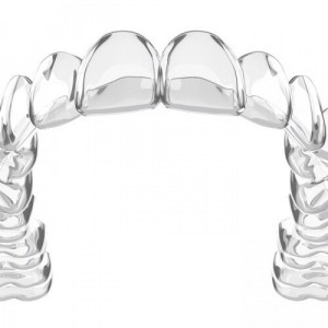
Aligners F22® is an Edra professional dentistry reference focused on clinical practice, education and treatment planning.
Products 10 July 2026
Planmeca introduces two dental units designed to support modern clinical workflows through different concepts and use cases.
News 10 July 2026

BIOLASE, a MegaGen company and global leader in dental laser technology, today announced a significant investment in its Corona, California, manufacturing operations, underscoring
News 10 July 2026

Led by Farhad Boltchi, DMD, and Darin Dichter, DMD, the program provides a real-time look at the clinical decisions that shape anterior implant outcomes


Copyright © 2026 - All Rights Reserved
ISSN 2767-1178