

Foreign body in the maxillary sinus
Authors: M. Gobbo, G. Ottaviani, K. Rupel, M. Biasotto
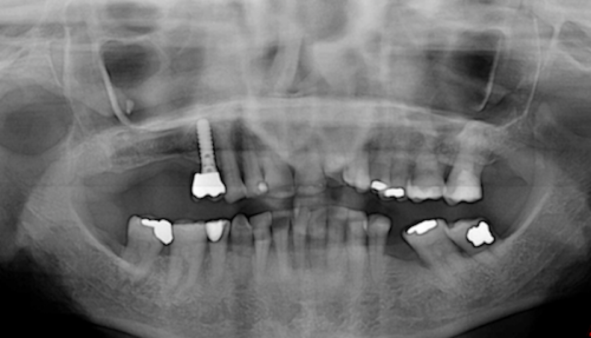
A female patient was sent to the Medicine and Oral Pathology clinic of the Dental Clinic of Trieste for the evaluation of an intensely radiopaque foreign body present in the right maxillary sinus. The patient had never had any symptoms. However, during a first visit performed at the Dental Clinic of Trieste, to which she presented herself with a recent orthopantomography (fig. 1a), the dentist had considered it appropriate to refer her to the Medicine and oral pathology clinic for further study, view also the intense inflammatory reaction visible radiographically. The patient, in good health, did not take any medication and only related to being allergic to tramadol.The right maxillary arch appeared partially edentulous, with the absence of elements from 1.4 to 1.8 and the presence of an osseointegrated implant in the edentulous area 1.4. During the first visit in oral pathology an intraoral X-ray was taken at site 1.4, from which no lesion was found. Clinically, the edentulous area was rosy, without continuous solutions or injuries. Given the clinical situation, age and good health of the patient, it was decided to intervene with the surgical removal of the foreign body under local anesthesia.
DIAGNOSIS AND CARE
Three weeks later the maxillary sinus was revised endoscopically. After written informed consent from the patient, local anesthesia was performed and a full-thickness trapezoidal flap was set up in zone 13-17. Subsequently, a bone trap was made with piezoelectric inserts, (figs. 2a, b), the Schneider membrane was incised and a thin and flexible fibroscopic probe was introduced to inspect the right maxillary sinus. Once the foreign body is highlighted, it has been removed using a scollatore and surgical tweezers (fig. 2c). The inflammatory reaction, which is visually polypoid, has always been removed and cured endoscopically (Fig. 3). The maxillary sinus was finally cleansed with the alternate use of sterile water and rifamycin. The bone trap created for access to the maxillary sinus was repositioned with mini 5 mm osteosynthesis plates (fig. 2d) and, subsequently, protected with lyophilized native collagen and with the repositioned flap. The patient was prescribed antibiotic therapy (amoxicillin 3 g / day for 6 days; N-acetylcysteine 300 mg / 3 ml to be taken every night with an aerosol). After a week, the silk sutures were removed. The patient denied pain, swelling or other symptoms. After 8 days the histological examination confirmed the presence of an intense phlogistic reaction and a lesion of likely mycotic origin, also called fungus ball. After 4 months, the picture appears stable. Sinusitis of fungal origin is a phenomenon in continuous growth. It is divided into two categories: invasive and non-invasive. Among the "non-invasive", there is an allergic form and the so-called fungus ball. While the first form is found as a consequence of hypersensitivity to allergens of fungal origin mediated by the naso-sinus tract, the fungus ball type is found in immunocompetent non-allergic subjects and seems to be linked to the accumulation of debris and fungal components in the paranaxal sinuses, usually from the Aspergillus family. Although they are initially asymptomatic pathologies, the long-standing fungus ball may present with non-specific symptoms such as nausea congestion, purulent or blood drainage from the ipsilateral nostril, headache, facial pain or alteration of the sense of smell. The pathogenesis of these lesions remains debated. The treatment of choice remains surgery with removal of the foreign body and revival of the affected breast, without any need for adjuvant antifungal therapy

Fig. 1 Preoperative orthopanoramic radiography showing the presence of the fungus ball in the right maxillary sinus (a). Post-operative radiograph showing the healing of the right maxillary sinus and the presence of osteosynthesis plaque (b)

Figs. 2a-d Access hatch construction to the maxillary sinus (a, b). Removal of the fungus ball (c). Repositioning of the bone trap and fixation with osteosynthesis screws (d)

Fig. 3 Aspect of the fungus ball through the endoscopic screen (image obtained with flexible probe) and polypoid inflammatory aspect of the right maxillary sinus affected by asymptomatic chronic sinusitis and presence of fungus ball
 Related articles
Oral pathology 02 June 2026
This peer-reviewed oral pathology article summarizes clinical evidence from Oral oncology (2026). It focuses on findings that may help dental professionals evaluate treatment decisions, patient...
Oral pathology 27 May 2026
This peer-reviewed oral pathology article summarizes clinical evidence from BMC oral health (2024). It focuses on findings that may help dental professionals evaluate treatment decisions, patient...
Oral pathology 26 May 2026
This peer-reviewed oral pathology article summarizes clinical evidence from Oral oncology (2026). It focuses on findings that may help dental professionals evaluate treatment decisions, patient...
Oral pathology 18 May 2026

This peer-reviewed oral pathology article summarizes clinical evidence from Oral oncology (2026). It focuses on findings that may help dental professionals evaluate treatment decisions, patient...
Oral pathology 13 May 2026

This peer-reviewed oral pathology article summarizes clinical evidence from BMC oral health (2024). It focuses on findings that may help dental professionals evaluate treatment decisions, patient...
Read more

New initiative invites dentists to experience DEXIS’ most advanced AI yet, built on scale, speed, and clinical trust.
News 05 June 2026

(Nasdaq: ALGN), a leading global medical device company that designs, manufactures, and sells the Invisalign® System of clear aligners, iTero™ intraoral scanners, and exocad™ CAD/C
News 05 June 2026

(Nasdaq: HSIC), the world’s largest provider of health care solutions to office-based dental and medical practitioners, today announced that its Board of Directors has elected Will

As the University of Colorado School of Dental Medicine celebrates the graduating DDS Class of 2025, we are proud to recognize the students and faculty members whose exceptional de
Oral surgery 05 June 2026
This peer-reviewed oral surgery article summarizes clinical evidence from International journal of oral and maxillofacial surgery (2026). It focuses on findings that may help dental professionals...


Copyright © 2026 - All Rights Reserved
ISSN 2767-1178