

Papillomatous gingival lesion
Authors: Giulio Villa, Elena Maddalone, Elena Maria Varoni, Davide Costa, Alberto Pispero, Laura Moneghin
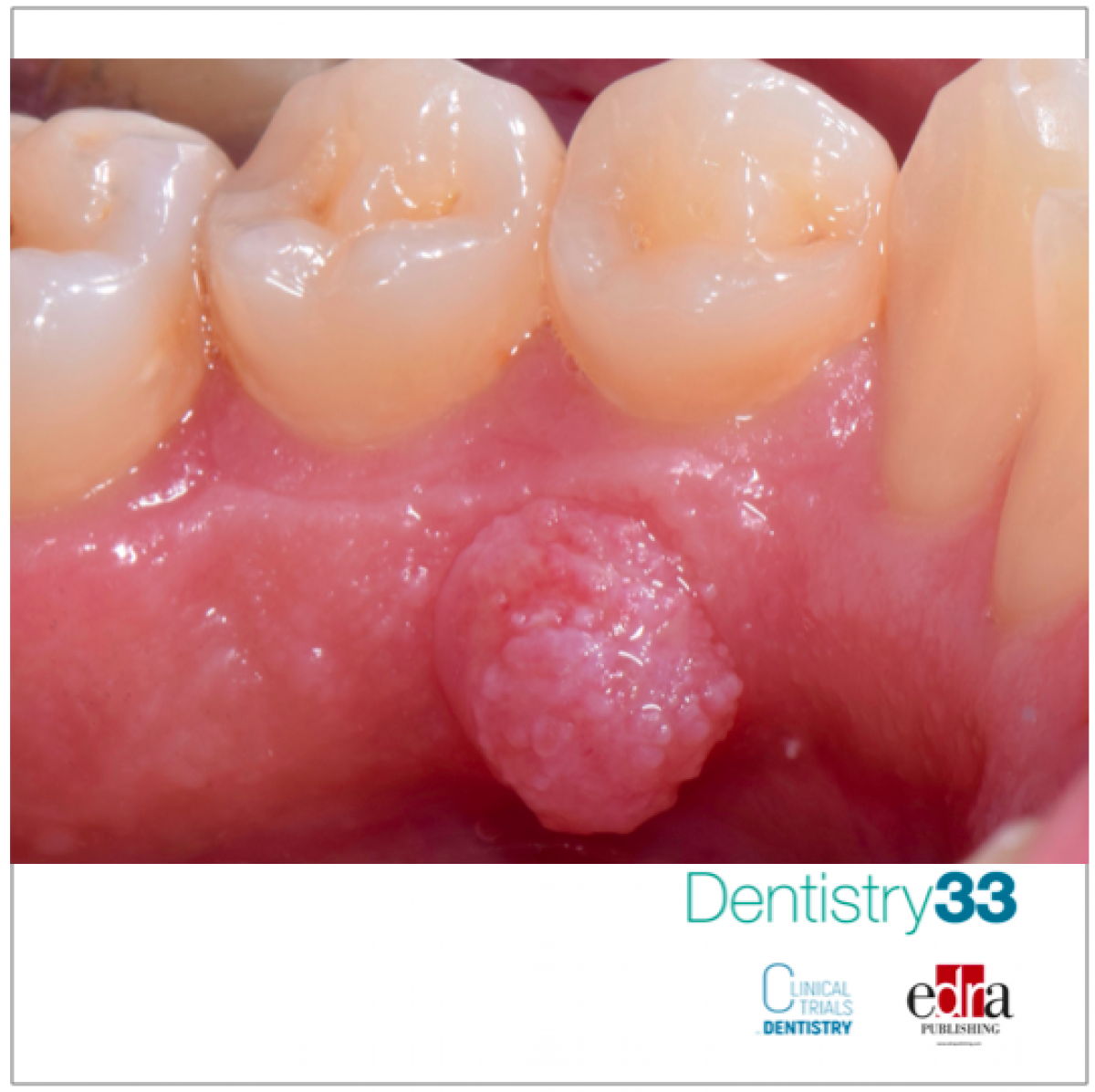
The patient, a former smoker, had a history of cardiac infarction which occurred about 3 years earlier, operated with the placement of 4 cardiac stents and was also suffering from arterial hypertension. The drug history reported the intake of low-dose acetylsalicylic acid, atorvastatin, patoprazole, bisopolol and amlodipine. The physical examination revealed the presence of a lesion in the gingiva adhering to the lingual aspect of the III quadrant, corresponding to elements 3.3-3.4. The lesion was exophytic, papillomatous, with a sessile implant base, soft consistency, pinkish-whitish in color, painless to palpation and with a diameter of about 1 cm.
Based on the clinical features highlighted during the physical examination, the lesion was placed in a differential diagnosis with squamous papilloma, pyogenic granuloma, squamous cell carcinoma and peripheral odontogenic fibroma. Subject to informed consent and local periwound anesthesia with 2% mepvacain with vasoconstrictor, an incisional biopsy was performed with the "cold blade" method and diamond incision. Hemostasis of the wound was achieved by compressing the intervention site with gauze soaked in physiological tissue and it was not necessary to apply sutures. The collected material was oriented on paper, immersed in fixative liquid and was sent to fellow anatomopathologists for microscopic analysis of the samples. Histopathological analysis yielded a proliferation result of the peripheral / extraosseous ameloblast type, worthy of radicalization. Ameloblastoma is a benign neoplasm of odontogenic epithelial origin that can present with various histological growth patterns. Referring to the definition of the World Health Organization (4th edition, 2017), ameloblastomas are classified as follows: solid / multicystic, peripheral / extrosseous, desmoplastic and unicistic. Currently, the classification has been simplified and restricted to ameloblastoma, unicistic ameloblastoma and peripheral / extraosseous type. The WHO defined peripheral ameloblastoma as "the extra-osseous counterpart of solid / multicystic ameloblastoma". Peripheral meloblastoma is thought to be the rarest subgroup, constituting 1 to 5% of all ameloblastomas, with a slight predilection for males and occurring most frequently in fifth to sixth decade of life. Clinically, it appears as an exophytic sessile mass, generally asymptomatic, with a smooth or papillary surface. It commonly affects the mandible, particularly the lingual gingiva in the premolar region, followed by the anterior region. However, there have been reports of extra-gingival positions, such as the buccal mucosa and the oral floor. There is rarely any radiological evidence of bone infiltration, however re-absorption of the underlying bone tissue may occur due to the pressure exerted by the tumor. The current therapeutic recommendation for peripheral ameloblastoma is complete surgical excision with adequate disease-free margins. Rare cases of relapse have been reported, even several years after surgery, progression of the disease and transformation of the disease, therefore a long-term follow-up is mandatory.
 Read more
Edra Professional Books 06 June 2026
Understanding Dental Insurance: A Practical Reference for Dental Professionals

Understanding Dental Insurance is an Edra professional dentistry reference focused on clinical practice, education and treatment planning.

New initiative invites dentists to experience DEXIS’ most advanced AI yet, built on scale, speed, and clinical trust.
News 05 June 2026

(Nasdaq: ALGN), a leading global medical device company that designs, manufactures, and sells the Invisalign® System of clear aligners, iTero™ intraoral scanners, and exocad™ CAD/C
News 05 June 2026

(Nasdaq: HSIC), the world’s largest provider of health care solutions to office-based dental and medical practitioners, today announced that its Board of Directors has elected Will

As the University of Colorado School of Dental Medicine celebrates the graduating DDS Class of 2025, we are proud to recognize the students and faculty members whose exceptional de


Copyright © 2026 - All Rights Reserved
ISSN 2767-1178