

Evaluation of periapical surgery under operative microscope: a retrospective study
Authors:Christian Bacci, Luca Donolato, Alessia Cerrato, Gastone Zanette

Apical post-treatment pathology can jeopardize the success of endodontic treatment. If periapical pathology develops the orthograde retreatment is considered the treatment of choice whenever possible, in adjunction to the surgical endodontic retreatment or tooth ex- traction in case of failure of the previous treatments. Aim of the present retrospective study was to evaluate the efficacy of periapical surgery using an endodontic microscope unit, at two years of follow up.
MATERIALS AND METHODS
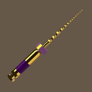
A retrospective cross-sectional study was conducted on 42 teeth from 36 patients. All standardized surgical procedures were performed using an operating endodontic microscope unit (magnification range: 5X-40X). The criteria to state success or failure were both clinical (swelling, pain, tooth tender to palpation) and radiographical (healing and ossification). In order to evaluate the healing of periodontal tissues, several control radiographs were performed at 1, 3, 6, 12, 24 months after the intervention.
RESULTS
During the 2-years follow up, sixteen teeth were lost (ten patients had not attended the recall examination, and two teeth had been extracted for other reasons; drop-out rate of 38%). The results of the present study showed that the postoperative heal- ing course was uneventful for 26 teeth. At the 1-year follow-up examination, 25 teeth (96%) showed no adverse clinical manifestations (clinical score = 0). Only one tooth was tender to vestibular palpation (clinical score = 1). Excellent results were also noted for the patients’ pain assessment, with 24 teeth (92%) exhibiting no painful symptoms (pain score = 0). Only 2 patients complained about a temporary painful condition (pain score = 1). After 1 year, 23 teeth (81%) showed radiographically a complete osseous healing (regeneration ≥90%) and 3 teeth (11%) exhibited an osseous regeneration between 50% and 90%.
DISCUSSION
According to the results of the present study, the authors underlined that radiographic criteria are useful component in deter- mining whether or not treatment is successful although symptom evaluation, as well as an accurate examination are critical determinants to discriminate between success and failure. Furthermore, the results of this study suggest that using the intraoperative microscope better results can be achieved, mainly due to a proper intraoperative diagnosis allowed by the use of the magnification unit. In other words, the microscope highlights the presence of complications, such as root fractures or perforations; thus, the apicectomy surgical treatment is completed only when it is clinically indicated.
CLINICAL SIGNIFICANCE
Based on the data from this retrospective study using an endodontic intraoperative microscope better results can be achieved, mainly due to a proper intraoperative diagnosis allowed by the use of the magnification unit. The microscope can, in fact, high- light the presence of complications, such as root fractures or perforations and consequently the apicectomy surgical treatment is completed only when it is clinically indicated.
 Tag
Tag
Related articles
Oral surgery 04 July 2022
Evaluation of the effectiveness of apicoectomy performed with the microscope: a retrospective study

Authors: Christian Bacci, Luca Donolato, Alessia Cerrato, Gastone Zanette
Apical post-treatment pathology can jeopardize the success of endodontic treatment. Various alternatives must be taken into account to promote periapical healing: orthograde retreatment, which is...
Read more
Products 26 June 2026
Coronal flaring is considered a key step in efficient root canal preparation, especially for difficult-to-access canals.
News 26 June 2026
The Association for Dental Safety (ADS) proudly announced the recipients of the 2026 Leadership Awards during its Annual Conference on May 27 in Salt Lake City, Utah.
News 26 June 2026

Recognition highlights the company’s doctor-led culture and continued investment in team member growth and engagement
Editorials 26 June 2026
From California to Canada: CU Anschutz School of Dental Medicine at ADEA, GRC and IADR 2026
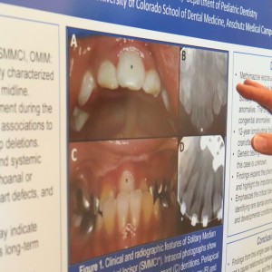
The CU Anschutz School of Dental Medicine will showcase a wide breadth of educational innovation, faculty development, clinical training and research at three major gatherings this
Oral surgery 26 June 2026
This peer-reviewed oral surgery article summarizes clinical evidence from International journal of oral and maxillofacial surgery (2026). It focuses on findings that may help dental professionals...


Copyright © 2026 - All Rights Reserved
ISSN 2767-1178