

3D facial scanners or 2D images for patient communication and treatment planning?
Edoardo Mancuso
Intraoral scanners, facial scanners, and computer-aided design software programs have become powerful tools for treatment planning and previsualization, and are everyday more often used by dentists in their practice.
The objective of incorporating digital technologies such as facial and intraoral scanners and CAD software programs might help improve patient communication and facilitate interdisciplinary dialogue.
Moreover, digital technologies facilitate the superimposition of 3D digital waxing into the 2D or 3D image of the patient, which represents a convenient diagnostic tool and may improve patient visualization of the outcome of the proposed treatment.
However even if digital previsualization might be a powerful tool, it has been reported that patients detect discrepancies in smile characteristics and esthetics, differently than dentists do, and this can undermine patients' satisfaction at the end of the dental treatment.
On this matter, Dr. Revilla-León has written an interesting observational study, recently published in the journal of prosthodontics with the aim of measure differences in esthetic perception among laypersons, dental students, and dentists. The objective is to assess the differences perceived by the subjects between the occlusal plane and dental midline discrepancies on a full-face photograph (2D image) or a digitized face (3D video).
Material and methods
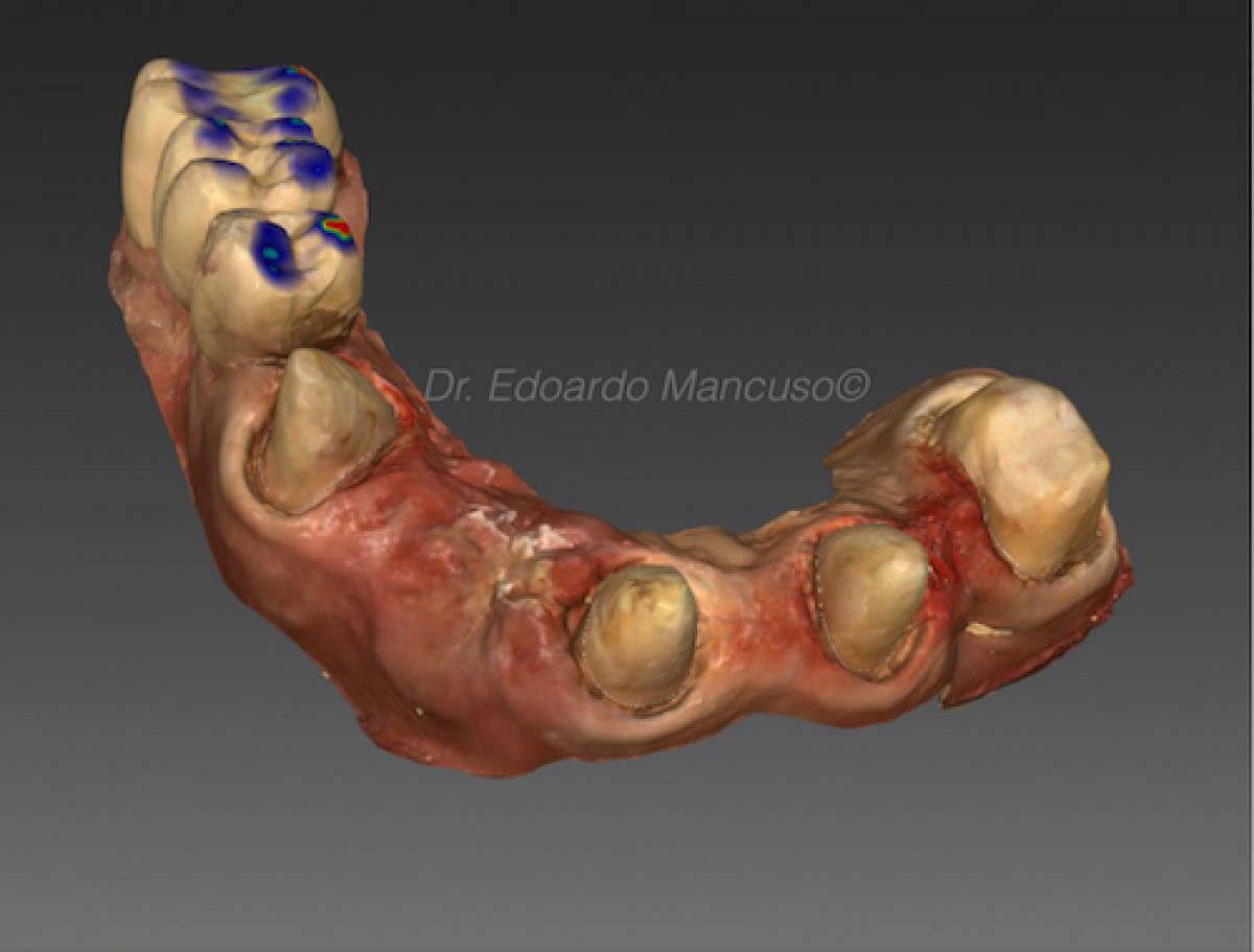
For the material and methods used in the article, a female model was digitized by using a facial scanner, intraoral scanner, and a full-face smile photograph. Dental discrepancies were simulated by using a 2D photograph and a 3D scan of the model. In both simulation groups, 2 subgroups were produced. The occlusal plane of the first subgroup was modified in 1-degree increments without changing the dental midline or the position of the maxillary dental incisors. In the second subgroup, the occlusal plane was modified by using the same increments, but the maxillary central incisors and dental midline were altered to match the inclination of the occlusal plane. A total of 300 participants were then asked to rate the 2D images and 3D videos on a 1-to-6 scale and answer a questionnaire. Ordinal logistic regression was used to analyze the ratings.
Results
For the results of the study, the layperson group gave consistently higher ratings than the other 2 groups in both the 2D and 3D simulations. The ratings decreased with the increased tilt of the occlusal plane. The rating decrease was higher for dentists, followed by students, and lastly by laypersons.
The use of 3D versus 2D images had a positive effect on the ratings, but the effect decreased for the student observers and decreased even further for laypersons. Furthermore, midline alteration led to higher ratings but also resulted in worsening of the odds ratio for the tilt. Seventy percent of the dentists, 57% of the dental students, and 52% of the laypersons preferred 2D simulations to 3D simulations.
Discussion
The study reports that dentists, dental students, and laypeople rated differently the disparities between the occlusal plane and the interpupillary and commissural lines with or without the alteration of the maxillary dental midline inclination on both 2D and 3D simulations. In the study, small perception differences were encountered on 3D simulations between dentists and dental students, with dental students providing lower ratings than dentists at the same occlusal plane tilt. Dr. Revilla-León writes that this could be explained by a generational difference or earlier adaptation of the technology on a daily basis.
The research resulted in a significant preference of the 2D simulations to the 3D from dentists, students, and laypersons. The article motivates this result explaining that evaluating the entire face and personality of a patient is extremely important when planning a treatment with extensive restorations. Psychological factors and subjective preferences of the patient should be also considered and incorporated into the treatment planning procedures. For these reasons the 3D representations may eliminate the emotional aspect, while the 2D photograph maintains the emotional component as participants may identify themselves better in a conventional photograph.
Conclusions
Based on the findings of the observational study, the following conclusions were drawn by the authors:
1. Dentists, dental students, and laypersons decreased their ratings with an increased inclination of the occlusal plane. Observer group ratings gradually diverged from the layperson group, giving consistently higher ratings than the dentists and dental students.
2. Overall, 3D simulations obtained higher ratings than 2D images in dentists and dental student populations, but the positive effect was reversed at higher occlusal plane tilt angles.
3. Laypersons graded all the 2D and 3D images as esthetically pleasant.
In conclusion, we can say that incorporating digital technologies such as facial and intraoral scanners and CAD software programs into private practice might help with patient communication, facilitates interdisciplinary dialogue, and improves diagnostic waxing. However, training and practice are needed to visualize 3D representations of patients, and even though 3D representation may provide a powerful diagnostic tool for the clinician, patients may not be able to interpret and visualize the 3D simulation appropriately. Consequently, a more accessible and understandable technology may be recommended based on the goal of the simulation.
For additional information: Esthetic dental perception comparisons between 2D- and 3D-simulated dental discrepancies
 Tag
Tag
Related articles
Periodontology 18 December 2025

Using the periodontal diseases classification published in 2018, this study evaluated the level of agreement among predoctoral and postgraduate students of different education levels and specialties...
Orthodontics 16 December 2025
Diagnosis and treatment planning of orthodontic patients with 3-dimensional dentofacial records

Cephalometrics has been the foundation of orthodontic diagnosis for many years. However, for many orthodontic patients, a lateral cephalogram might not be necessary.
Implantology 17 October 2025
To investigate whether cross-section imaging influences the planning and therapy of standard implant cases in the posterior mandible.
Endodontics 16 October 2025
Endodontic or dental implant therapy: The factors affecting treatment planning
Clinicians are confronted with difficult choices regarding whether a tooth with pulpal and/or periapical disease should be saved through endodontic treatment or be extracted and replaced with an...
Digital Dentistry 15 October 2025
The Impact of Artificial Intelligence on Diagnostic Accuracy and Treatment Planning in Dentistry

The use of AI in dentistry is revolutionizing the field of dentistry by enhancing the accuracy of diagnosis and treatment.
Read more

Eagle Crown Lengthening Burs are designed to make surgical precision effortless—helping clinicians expose more tooth structure smoothly, efficiently, and with total control.
News 17 July 2026

The new Bogotá, Colombia facility strengthens Roland DGA’s long-standing commitment to dental professionals and partners across the region.

New integration streamlines patient financing within CareStack’s practice management platform, making it easier for providers to help patients move forward with care.

As the University of Colorado School of Dental Medicine celebrates the graduating DDS Class of 2025, we are proud to recognize the students and faculty members whose exceptional de
Oral surgery 17 July 2026

This peer-reviewed oral surgery article summarizes clinical evidence from International journal of oral and maxillofacial surgery (2025). It focuses on findings that may help dental professionals...


Copyright © 2026 - All Rights Reserved
ISSN 2767-1178