

Soft tissue surgery at implant abutment connection for improving aesthetics: 6-month outcomes of a multicentre randomised controlled trial of xenogeneic dermal matrix versus autologous connective tissue graft versus no graft
Authors: Niccolò Baldi, Jacopo Buti, Magda Mensi, Fortunato Alfonsi, Chiara Cinquini, Paolo Tonelli, Antonio Barone

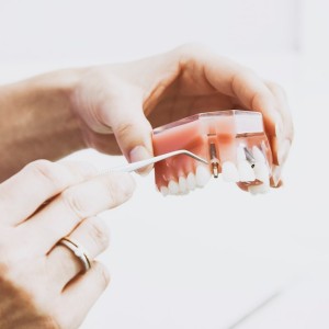
Objectives. The aim of this multicentre randomised controlled trial was to evaluate the efficacy of a xenogeneic dermal matrix in widening keratinised peri-implant tissues during second-stage surgery, and to compare it to both autologous connective tissue graft and a control group with no augmentation.
Material and methods. Patients requiring an increase in keratinised gingiva width were enrolled by four university/dental practices and randomised into three different groups for grafting procedures at the implant uncovering stage: either xenogeneic dermal matrix (Group X), autologous connective tissue graft (A) or no graft (control, Group C). The primary outcomes were width of keratinised tissue and facial soft tissue levels, evaluated at three different time points (T0, implant uncovering stage; T1 and T2, six weeks and six months after surgery, respectively). Secondary outcomes were: implant failure, complications, marginal bone loss, papilla index, facial soft tissue level, pink aesthetic score, and aesthetic assessment by patients.
Results. Thirty-six patients, with one implant per patient, were enrolled at two centres (18 at each centre): 12 for control, 12 for xenogeneic dermal matrix and 12 for autologous tissue graft. Three patients dropped out and two patients from the autologous group had implant failures. No complications were recorded. After six months, the width of keratinised tissue increased by 0.16 ± 1.01 (P = 0.79), 1.05 ± 0.76 (P = 0.01) and 0.80 ± 1.73 mm (P = 0.28), and facial soft tissue level was -0.95 ± 0.85 (P = 0.04), 0.32 ± 0.57 (P = 0.15) and 0.35 ± 0.79 mm (P = 0.30) respectively in Groups C, X and A groups. Between-group analysis showed that, with respect to control, only facial soft tissue level (1.31 mm, P = 0.01) and width of keratinised mucosa (2.43 mm, P = 0.01) outcomes in the autologous graft group were statistically significant at T2. Mean marginal bone loss between T0-T2 was -0.4 ± 0.4mm, with no differences between groups. Pink aesthetic score showed no significant differences between groups, being 0.89 for A-C (P = 0.41), 0.88 for A–X (P = 0.63) and 0.72 for X-C (P = 0.88).
Patient’s aesthetic satisfaction (Visual Analogue Scale) was 92.2 ± 8.4, 93.8 ± 7.7, 97.2 ± 3.0, for Groups C, X and A, respectively. Between the two dental centres, only facial soft tissue level at T0–T2 was significantly different, by 0.67 ± 0.62 mm (P = 0.03).
Conclusions. After six months, autologous connective tissue graft yielded a significant gain in facial soft tissue levels and width of keratinised mucosa, as compared to the control group (no graft).
 Tag
Tag
Read more
Products 24 July 2026
Expanded shade range supports fast, esthetic chairside zirconia restorations with 9-minute sintering when used with CEREC ® SpeedFire
News 24 July 2026

Sensei, a Carestream Dental brand and a global leader in dental practice management software and digital imaging solutions, today announced a strategic partnership with MAX Surgica
News 24 July 2026

Beach Point Capital Management LP (“Beach Point”), a global alternative investment manager with over $20 billion in assets under management, today announced the successful sale of
Editorials 24 July 2026
Achievements Unlocked: DDS Class of 2024 Student and Faculty Awards Announced

Rick Mediavilla, DDS, FAGD, associate professor and associate dean of admission, student and alumni affairs, had the honor of announcing each of the 79 graduates, their plans follo
Implantology 24 July 2026
This peer-reviewed implantology article summarizes clinical evidence from Clinical oral implants research (2026). It focuses on findings that may help dental professionals evaluate treatment...


Copyright © 2026 - All Rights Reserved
ISSN 2767-1178