

Zygomatic Implant Guide “ZIG” A New guide-plate concept.
Authors: F. Zingari F. Gallo S. Ghezzi E. Carozzi

Zygomatic implantology has made significant progress over the past 20 years. Initially, it defined success rates, in terms of survival, comparable to those of traditional implants, (1-3) over time, it has increasingly oriented its scientific aim to the reduction of post-surgical problems. (4-5) Currently, zygomatic implantology is aiming at improving its standards of precision and surgical-prosthetic predictability. (6) In this sense, surgery is having great development
guided zygomatic. (7-9)
Clinical question
In the past, various techniques of guided zygomatic surgery have been described; (10) however, the complexity of the surgical site on the one hand and the sophistication of the proposed guided techniques on the other (double plates, closed bushings, mucous-supported plates), did not allow the affirmation of a true guided surgery technique that it was, at the same time, precise and easy to use. Specifically, in fact, the past techniques of guided surgery on zygomatic implants were nothing more than elaborations of the guided technique on traditional implants, with the consequent difficulty, if not impossibility, of using the various drills in a single surgical step and following the procedure. zygomatic implant itself in a “full-quided” manner How to obtain a zygomatic surgery technique full-guided precise and easy to use? An innovative full-quided zygomatic surgery technique that solves the specific problems of the previous guided techniques will be described below.
Description
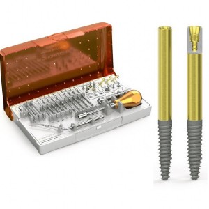
3 clinical cases were included in the study: in the first case, the surgical project involved the insertion of 2 zygomatic implants and 4 traditional premaxilla implants (4 BLT Straumann implants diameter 4.1 x 12 mm in length and 2 zygomatic implants Noris Medical diameter 4.2 by 50 mm in length), in the second case, the surgical project involved the insertion of 4 zygomatic implants and 2 traditional premaxilla implants (number 2 Neodent Alvim implants with a diameter of 3.5 by 10 mm in length and number 4 Noris Medical zygomatic implants diameter 4.2 for lengths 42.5; 47.5: 37.5 and 52.5 mm), in the third case, the surgical project involved the insertion of 4 zygomatic implants, 2 pterygoid implants and 1 implant traditional in premaxilla (number 4 Noris Medical zygomatic implants diameter 4.2 mm by length 35 mm and three 47.5 mm number two pterygoid Noris Medical diameter 4.2 mm by length 20 mm, number 1 Neodent Alvim implant diameter 3.5 by 10 mm of lu length). The technique was used in all clinical cases "Zygomatic Implant Guide" (ZIG), therefore, patients underwent a massive facial CT scan (or CBCT). The radiographic examination expressed in Dicom files is imported into the design software and associated with the STL files obtained from a scan of the project prosthetic. In the first case, the prosthetic design associated with the sufficient amount of residual bone in the premaxilla allowed us to insert 2 zygomatic implants in the area 26 and 16 and to place 4 traditional implants in the premaxilla area (Figs. 1,2).

In the second case, the prosthetic project associated with the amount of residual bone suggested us to insert 4 zygomatic implants in the 14-16-24-26 area and 2 traditional implants in premaxilla (Figs. 3,4)

In the third case, the prosthetic project associated with the amount of residual bone suggested us to insert 4 zygomatic implants in the area 14-16-24-26, 2 pterygoid implants in the area 17 and 27 and 1 traditional implant in the subspinal region (Figs. 5.6 ). In order to produce the surgical guide (ZIG), the position and length information of the chosen implants is transferred to a second software for the CAD design.
In this CAD drawing phase, the shape of the plate and the position of the bushings are defined, which will determine both the subsequent drill steps and the insertion of the zygomatic implants.
The shape of the plate is designed to fully support the bone and is fixed with screws or pins so as to be stable during all phases of use (Fig. 7).
The precision of the plate and its bone fitting are crucial, in order to obtain a predictable result, therefore, it is essential to perform high definition CT / CBCT and produce the "ZIG" with the Laser-Melting technique. The uniqueness of the plate, protected by a registered patent, consists in not having the common access bushings for the instruments (drills and implants). The closed bushings used up to now, in fact, force the surgeon to perform extremely complex and invasive maneuvers, often not tolerated by the patient himself. In fact, the insertion of the posterior implant (implant to be inserted in any case of zygomatic implantology) was extremely complex, if not impossible, due to the reduced buccal opening of the patients in that specific area of the oral cavity.
Thanks to the "open" bushing technique, consisting of a half-bushing and a counter-half-bushing (Fig. 8), the problem of the overall dimensions of the instruments in the oral cavity is solved and, therefore, it is possible to proceed with all the drill steps and, even , insert the implant under full guidance of the plate. The plate is then removed only after inserting the implant as per the presurgical program, making the entire surgical act "full guided". Biomechanically, the half-bushing and counter-half-bushing define a common longitudinal median axis; called half-bushes, having a semicircle section with opposing concavities, they define a seat in which the instruments (drill and implant)
they work in a completely guided way according to a predetermined axis.


Surgical technique
Clinical cases were treated under local anesthesia with articaine + adrenaline 1: 100,000 (with the exception of the case of 4 zygomatics + 2 pterygoids + 1 anterior implant in which the patient, dental phobic, was operated on under general anesthesia). The incision was made 5 mm palatal to the alveolar ridge with a median and distal vestibular drain in correspondence of the tubers. A detachment of the soft tissues is performed in a gentle way subperiosteally, with preservation of the infraorbital nerves. Once the region was detached, the plates were positioned in such a way as to
obtain a full fitting with the receiving bone structure (Fig. 9). Plate fixation was perfected with micro screws (1.5 mm) (Fig. 10). The plates have, in correspondence with the counter-half-bushing, a guide rail for the first cutter which is represented by a diamond ball cutter (Fig. 11). After identifying the drilling point, thanks to the spot impressed in the bone by the diamond ball, we proceed with the use of a cylindrical diamond bur able to remove, always under the absolute guide of the plate, all the bone quantity from the surface of the maxilla and cheekbone, which would interfere with the passage of the drill first and then the implant (Fig. 12). Both the passage of the zygomatic bone drilling drill (Fig. 13), and the subsequent insertion of the implant (Fig. 14), therefore take place using the template and without the patient has to make forced buccal openings or that the surgeon has to perform articulated maneuvers. The correct insertion of the implants followed positioning of the MUAs to correct implant disparallelism and immediate loading. 11,12 After 4 months, all clinical cases were finalized with a definitive prosthesis.


Conclusions and clinical indications
Zygomatic implantology, although an object of ever-increasing scientific interest, has not yet established itself in clinical dental practice, due to the difficulty of the anatomical site and the complexity of the implant-prosthetic treatment. The ZIG technique
offers the implantologist the simplification of the surgical procedure, the predictability of the result and the complete respect of the prosthetic project. The use of the ZIG technique is potentially usable in any case of severe maxillary atrophy that requires zygomatic implantology.

Bibliografia
1. Boyes-Varley JG, Howes DG, Lownie JF, Blackbeard GA. Surgical modifications to the Bränemark zygomaticus protocol in the treatment of the severely resorbed maxilla: a clinical report. Int J Oral Maxillofac Implants. 2003:18:232-237
2. Galán Gil S, Peñarrocha Diago M, Balaguer Martínez J, Marti Bowen E. Rehabilitation of severely resorbed maxillae with zygomatic implants: an update. Med Oral Patol Oral Cir Bucal. 2007:12:E216-220
3. Bothur S, Jonsson G, Sandahl L. Modified technique using multiple zygomatic implants in reconstruction of the atrophic maxilla: a technical note. Int J Oral Maxillofac Implants.2003:18:902-904.
4. Molinero-Mourelle P, Baca-Gonzalez L, Gao B, Saez-Alcaide LM, Helm A, Lopez-Quiles J. Surgical complications in zygomatic implants: A systematic review. Med Oral Patol Oral Cir Bucal.2016:21:e751-e757.
5. D'Agostino A, Trevisiol L, Favero V, Pessina M, Procacci P, Nocini PF. Are Zygomatic Implants Associated With Maxillary Sinusitis? Oral Maxillofac Surg. 2016;74:1562-1573.
6. Grecchi F, Bianchi AE, Siervo S, Grecchi E, Lauritano D, Carinci F. A new surgical and technical approach in zygomatic implantology. Oral Implantol (Rome). 2017;10:197-208.
7. Schiroli G, Angiero F, Silvestrini-Biavati A, Benedicenti S. Zygomatic implant placement with flapless computer-guided surgery: a proposed clinical protocol. J Oral Maxillofac Surg. 2011:69:2979-2989.
8. Van Steenberghe D, Malevez C, Van Cleynenbreugel J, Bou Serhal C, Dhoore E, Schutyser F, Suetens P, Jacobs R. Accuracy of drilling guides for transfer from three-dimensional CT-based planning to placement of zygoma implants in human cadavers. Clin Oral Implants Res. 2003;14:131-136.
9. Wu YQ, Zhang ZY, Zhang CP, Huang W, Sun J, Zhang ZY. The installation of zygomatic implants and drilling guide. Zhonghua Kou Qiang Yi Xue Za Zhi. 2006 Mar; 41 (3):1 40-3.
10. ChowJ. A novel device for template-guided surgery of the zygomatic implants. Int J Oral Maxillofac Surg. 2016:45:1253-1255.
11. Tuminelli FJ, Walter LR, Neugarten J, Bedrossian E. Immediate loading of zygomatic implants: A systematic review of implant survival, prosthesis survival and potential complications. Eur J Oral Implantol. 2017:10:79-87.
12. Davó R, Pons O. 5-year outcome of cross-arch prostheses supported by four immediately loaded zygomatic implants: A prospective case series. Eur J Oral Implantol. 2015;8:169-174.
13. Chow J, Hui E, Lee PK, Li W. Zygomatic implants--protocol for immediate occlusal loading: a preliminary report. J Oral Maxillofac Surg. 2006;64:804-811.
 Read more
Products 24 June 2026
News 24 June 2026

The program celebrates graduates joining the nationwide Aspen Dental network in a landscape of growing demand for oral healthcare professionals.
News 24 June 2026
Mitsui Chemicals recently announced its intention to acquire Ultradent Products Inc., a global leader in cosmetic, preventive, and restorative dentistry.
Editorials 24 June 2026
Personal trauma leads to dental hygiene career for new Texas A&M College of Dentistry honor grad

Endodontics 24 June 2026
This peer-reviewed endodontics article summarizes clinical evidence from BMC oral health (2026). It focuses on findings that may help dental professionals evaluate treatment decisions, patient...


Copyright © 2026 - All Rights Reserved
ISSN 2767-1178