

Socket sealing post-extractive with xenogenic porcine collagen matrix: a prospective clinical trial
Co-authors: A. L. Rossi, V. Capilupi, D. Palombo - Department of biomedical, surgical and dental sciences, Università degli studi di Milano, ASST Santi Paolo e Carlo, Clinical Unit of Oral Surgery
Matteo Chiapasco

INTRODUCTION
Tooth extraction is associated with a significant alteration of the soft tissues surrounding the post-extraction alveolus. The alveolar bone crest preservation techniques are used in clinical practice to reduce the volumetric bone contraction that occurs as a result of the avulsion of a dental element and, at the same time, to restore the correct profile of hard and soft tissues. Within these techniques, different research activities have focused on the development of new materials and new surgical techniques, useful for the increase and preservation of the keratinized mucous tissue positioned around the rehabilitations on implants.
The objective of this prospective cohort study is to test the performance of a new xenogenic collagen matrix as a socket sealing material, to allow second-intention healing of post-extractive sockets filled with a xenogenic bone substitute or with an immediate submerged implant.
MATERIALS AND METHODS
10 patients were recruited, presenting a single-rooted tooth scheduled for extraction. After atraumatic tooth removal, the post-extractive alveolus received either a socket preservation procedure or an immediate submerged implant. In both cases, the gingival margins of the alveolus were sealed with a xenogenic collagen matrix (Mucoderm, Botiss Dental, Zossen, Germany). The following parameters were evaluated:
- exposed surface of the matrix at the end of surgery (T0);
- soft tissue healing at 1, 4, 6, and 8 weeks from surgery (T1-4);
- histological aspect of gingiva samples, harvested 20 weeks after surgery (T5);
- aesthetic performance provided by the socket sealing material (T4).
RESULTS
The results of the study showed that:
- the mean post-operative exposure area of the matrix was 26.25 mm2 (14.2 to 38.84 mm2 );
- 8 weeks after surgery, full wound closure was achieved in 9 out of 10 sites with healthy keratinized tissue;
- the histological evaluation of gingiva sample revealed the presence of healthy keratinized gingival tissue, with no signs of aberrations or anomalies;
- the mean colorimetric score ∆E between the regenerated site and the surrounding gingiva was 3.76 (3 to 6.55).
Seven out of 10 patients reported an excellent aesthetic integration of the matrix (∆E < 3.7).
CONCLUSIONS
Preliminary results from this study suggest that this new xenogenic porcine-derived collagen matrix could represent a valuable alternative to allow second intention healing of post-extractive sockets filled with a xenogenic bone substitute or with an immediate submerged implant. New randomized clinical trials are needed to confirm the preliminary results obtained in the study and evaluate the long-term benefits.
CLINICAL SIGNIFICANCE
The alveolar bone crest preservation techniques are used in clinical practice to reduce the volumetric bone contraction that occurs as a result of the avulsion of a dental element and, at the same time, to restore the correct profile of hard and soft tissues. In the present study, the use of the xenogenic matrix in collagen, in the coverage of a post-extraction site, seem to allow a correct regeneration and integration of the keratinized gingival tissue overlying the alveolus, both from a biological and an aesthetic point of view. However, the lack of consolidated scientific evidence regarding this technique requires the execution of new randomized clinical trials, aimed at confirming the results obtained, evaluating their long-term benefits.
For additional information:
Dental Cadmos n° 05/2018 - https://doi.org/10.19256/d.cadmos.05.2018.06

Fig. 1 Initial situation, clinical picture, frontal view. The element 2.1 results seriously compromised. We decided to proceed to its avulsion and then with implant-prosthetic rehabilitation.

Fig. 2 Initial situation, periapical intraoral radiography, insufficient root size and contraindication of element recovery

Fig. 3 Post-extractive alveolus; minimally invasive avulsion, frontal vision

Fig. 4 Biomaterial grafting into the alveolus; it will provide support for soft tissues above

Fig. 5 Post-extraction socket cover using the collagen matrix, adapted to the alveolar morphology and covered, for at least 2/3 of its surface, from the mucous flap, without periosteal incisions

Fig. 6 Stabilization of the mucosal flap and of the matrix through stitches in nylon 5/0. The size of the exposed matrix surface is highlighted

Fig. 7 Clinical check at 8 weeks from the intervention. Front view. In evidence the perfect aesthetic integration of the newly formed keratinized tissue with the surrounding tissue

Fig. 8 Clinical check at 8 weeks from the intervention. Occlusal view. The perfect aesthetic integration of the keratinized tissue is highlighted newly formed with the surrounding tissue
 Related articles
Oral surgery 17 July 2026

This peer-reviewed oral surgery article summarizes clinical evidence from International journal of oral and maxillofacial surgery (2025). It focuses on findings that may help dental professionals...
Oral surgery 08 July 2026

This peer-reviewed oral surgery article summarizes clinical evidence from International journal of oral and maxillofacial surgery (2026). It focuses on findings that may help dental professionals...
Oral surgery 03 July 2026

This peer-reviewed oral surgery article summarizes clinical evidence from International journal of oral and maxillofacial surgery (2025). It focuses on findings that may help dental professionals...
Oral surgery 26 June 2026
This peer-reviewed oral surgery article summarizes clinical evidence from International journal of oral and maxillofacial surgery (2026). It focuses on findings that may help dental professionals...
Oral surgery 18 June 2026

This peer-reviewed oral surgery article summarizes clinical evidence from International journal of oral and maxillofacial surgery (2026). It focuses on findings that may help dental professionals...
Read more
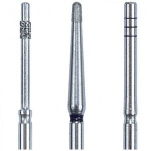
Eagle Crown Lengthening Burs are designed to make surgical precision effortless—helping clinicians expose more tooth structure smoothly, efficiently, and with total control.
News 17 July 2026
The new Bogotá, Colombia facility strengthens Roland DGA’s long-standing commitment to dental professionals and partners across the region.
New integration streamlines patient financing within CareStack’s practice management platform, making it easier for providers to help patients move forward with care.
As the University of Colorado School of Dental Medicine celebrates the graduating DDS Class of 2025, we are proud to recognize the students and faculty members whose exceptional de
Oral surgery 17 July 2026
This peer-reviewed oral surgery article summarizes clinical evidence from International journal of oral and maxillofacial surgery (2025). It focuses on findings that may help dental professionals...


Copyright © 2026 - All Rights Reserved
ISSN 2767-1178