

Gingival swelling adjacent to the dental elements roots: a clinical case
Marco Berardini

A male patient of 43 years comes to our observation, complaining about the presence of a roundish gingival excretion in the anterior area of the upper jaw. The patient reports that he has already noticed the lesion for a long time (a few years) and that he has not visited a specialist examination due to negligence and a subsequent depressive symptomatology. He also reports that he has noticed a slow but progressive increase in the size of the lesion in question.
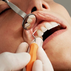
On intra-oral examination, the lesion appears as a swelling of diminutive consistency, of a pinkish color uniform, well bounded by surrounding healthy tissues and associated with elements 1.2 and 1.3 (figure 1). The size is about 1 cm and there are no signs of ulceration or purulent exudation. The intra-oral radiographic examination (fig. 2) also allows to exclude any involvement of the underlying hard tissues and, together with the clinical features, allows us to presume the absolute benignity of the lesion.
Obtained informed consent of the patient to treatment and given the clinical characteristics of presumed benignity (net margins, no bone involvement, soft consistency, uniform color, absence of ulceration and / or necrosis, clear delimitation with circus tissues - standing), we proceed with the total excision of the lesion with perilesional margins of 1 mm. The roots of the associated dental elements (1.2 and 1.3) are carefully cleaned and smoothed in order to remove the parodontal ligament cells which are likely to be at the origin of the lesion. The biopsy sample is immediately immersed in 4% formalin and sent to the laboratory for histological analysis (fig. 3). The periosteum around the area subject to removal is subdued with the blade of the scalpel in order to be able to pull the flap and avoid leaving the exposed bone tissue (fig. 4). The pathologist detects the presence of multinucleated giant cells distributed in an edematous and mucucinose stroma containing blood vessels and fibroblasts.
Based on the histological examination, a gigantocellular epulis is diagnosed. The etiology includes an irritative-inflammatory noxa that stimulates the abnormal proliferation (benign) of the cells of the periodontal bond. For complete recovery, treatment includes simple excision and removal of all local irritants.
For additional information:
Dental Cadmos n° 2/17 - https://doi.org/10.19256/d.cadmos.02.2017.04

Fig.1 - Evident roundish gingival lesion, on the intraoral objective examination, associated with elements 1.2 and 1.3

Fig. 2 - The intraoral radiography does not show signs of involvement of the underlying hard tissues to the injury

Fig. 3 - The lesion, completely removed, has a size of about 1 cm

Fig. 4 - Detached stitches. Horizontal cuts were made in the buccal periosteum to mobilize the flap as much as possible and to pull it in a coronal direction so as to leave a minimum amount of bone exposed
 Related articles
Oral pathology 13 July 2026

This peer-reviewed oral pathology article summarizes clinical evidence from Oral oncology (2026). It focuses on findings that may help dental professionals evaluate treatment decisions, patient...
Oral pathology 06 July 2026
This peer-reviewed oral pathology article summarizes clinical evidence from Oral oncology (2026). It focuses on findings that may help dental professionals evaluate treatment decisions, patient...
Oral pathology 02 July 2026

This peer-reviewed oral pathology article summarizes clinical evidence from BMC oral health (2024). It focuses on findings that may help dental professionals evaluate treatment decisions, patient...
Oral pathology 29 June 2026

This peer-reviewed oral pathology article summarizes clinical evidence from Oral oncology (2026). It focuses on findings that may help dental professionals evaluate treatment decisions, patient...
Oral pathology 19 June 2026
This peer-reviewed oral pathology article summarizes clinical evidence from BMC oral health (2024). It focuses on findings that may help dental professionals evaluate treatment decisions, patient...
Read more

The Sonicare brand’s first electric toothbrush powered by on-device AI and real-time spatially aware guidance helps patients brush with greater coverage and confidence for ultimate
News 15 July 2026

Young Innovations, a leading global manufacturer and distributor of dental supplies and equipment, announced the appointment of Rebecca Whitney as Chief Executive Officer, effectiv
For the second year in a row, the American Association of Orthodontists will celebrate Smiles at 7 Day on July 7.
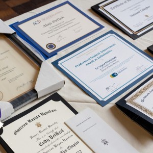
Achievement and service take center stage as the University of Colorado Anschutz School of Dental Medicine honors the Doctor of Dental Surgery (DDS) Class of 2026.
Oral pathology 15 July 2026

This peer-reviewed oral pathology article summarizes clinical evidence from BMC oral health (2024). It focuses on findings that may help dental professionals evaluate treatment decisions, patient...


Copyright © 2026 - All Rights Reserved
ISSN 2767-1178