

Lower lip mucosa neoformation
Giovanni Lodi
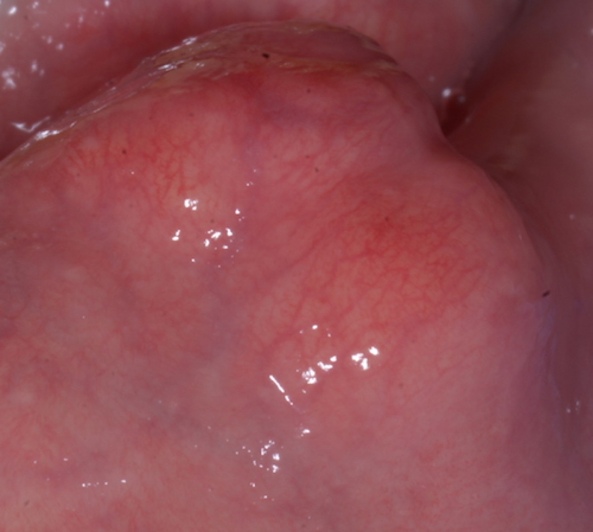
A 60-year-old patient in good general health is visited at the Oral Pathology and Surgery Service of the Operative Unit of Odontostomatology II of ASST Santi Paolo e Carlo. The medical history reveals mild arterial hypertension and type II diabetes mellitus being treated with Telmisartan 20 and Metformin 500 respectively. The non-smoker patient complains of the presence of a slow and continuous painless swelling at the mucous side of the left lower hemilab (fig.1 and fig.2). There are no skin changes in color or temperature, external palpation shows an elastic consistency in the area of the lesion, negative palpation of ipsilateral laterocervical lymph nodes.
At the intraoral level, a sessile swelling of about 2cm in diameter is observed, roundish, covered with a mucous membrane similar in appearance to the surrounding one and with a yellowish-pink color. On palpation the consistency is elastic and the lesion and the lesion is mobile below the mucous plane. No other alterations in shape or color of the intraoral mucous membranes are appreciated.
DIAGNOSIS AND CARE
The lesion on the basis of clinical data and that provided by the patient has characteristics of benignity. However, it is necessary to obtain a histological diagnosis that precisely determines the nature of the neoformation. The patient, after signing the informed consent, consents to the surgical treatment. Before proceeding with the biopsy intervention, a needle aspiration is performed at the center of the swelling through the mucous plane which does not show the presence of blood, salivary or similar purulent material. It is therefore possible to proceed with the intervention which in this case will be an excisional biopsy.
After perilesional infiltration of local anesthetic with vasoconstrictor, a mucosal incision of about 1cm is made above the lesion perpendicularly to the vermilion edge of the lip. The dissection of the tissues by blunt way through the primary incision allows to highlight a mass of frankly yellowish complexion. It is possible to easily cleave the lesion which appears frankly elastic, compact and separated from the surrounding tissues by a pseudocapsule of lining (fig. 3). At the end of the operation, the bottom of the operating field is clean and bloodless and the suturing of the incision margins takes place by means of a 5/0 absorbable polyfilament.
The diagnosis, confirmed by the histological examination, is of fibrolipoma (fig. 4). It is a benign tumor formed by a mix of fat and connective cells and is the most common histological variant of the lipoma. This pathology particularly affects limbs and trunk, when multiple sites are affected it is called multiple lipomatosis. At an intraoral level, it originates from the adipose and connective cells of the submucosa and clinically presents with a sessile or pedunculated yellowish pink swelling. The vestibular mucous membranes and the oral floor are mainly affected, following the tongue and lips. Diagnosis is simple considering the clinical aspects of color and consistency, slow growth and absence of symptoms. The differential diagnosis is with vascular lesions or from salivary tissues, due to obstructive problems of the duct or due to increases in the volume of the parenchyma. Needle spraying is a good way to distinguish these pathologies. The treatment indicated in the literature is the complete excision of the neoformation, an operation that is generally simple thanks to the ease with which it is possible to cleave the whole body of the lesion from the surrounding tissues. Relapses are very rare except for lipomas that infiltrate the muscles.

Fig1: intraoral aspect

Fig2: extraoral aspect

Fig3: lesion isolation

Fig4: histological image
 Related articles
Oral pathology 26 September 2023

The habit of smoking cigarettes has been associated with health problems for some time now, especially with regards to the link with diseases related to the...

Author: M. Berardini
A male patient, aged 60, underwent a visit complaining about the presence of a whitish and detected neoformation located in the right anterior abdomen of the tongue...
Oral pathology 06 August 2022

Co-authors: F. Scotti, M. Mandaglio, S. Decani, E. Baruzzi, L. Moneghini
An 85-year-old patient comes to our attention complaining of the presence of swelling of the right half-left, present for some time unspecified and gradually increased. The patient's medical history...
Oral pathology 01 August 2022

Author: Riccardo Mauro Bonacina
Patient AB, a 46-year-old male, comes to our observation complaining of an increase in volume at the palatal level for about a month. The systemic anamnesis...
Oral pathology 13 June 2022
The correlation between the quality of oral hygiene and oral HPV infection in adults

Aim of the study was to detect a possible association between the objectively determined quality of oral hygiene and the presence of oral HPV. MATERIALS AND METHODS In this perspective...
Read more
Digital Dentistry 18 April 2024


The Columbia University College of Dental Medicine (CDM) and the Fu Foundation School of Engineering and Applied Science have received approval from the New York State Department of Education to...

Dr. Richard Eidelson, DDS, MAGD, a nationally recognized leader in cosmetic dentistry, is thrilled to announce the opening of his second dental practice, Premier Dentist Philadelphia, located in the...

DDS Lab (DDS), one of the largest full-service dental laboratories in the world, today announced the commencement of operations out of its new state-of-the-art, full-service dental laboratory based...
d5.jpg)
The global electric toothbrush market size is estimated to grow by USD 2780 million from 2023 to 2027, according to Technavio. The market is estimated to grow at a CAGR of over 8.24% during the...


Copyright © 2024 - All Rights Reserved
ISSN 2767-1178